INTRODUCTION
AITP is an autoimmune disorder characterized by a low platelet count and mucocutaneous bleeding1). Splenectomy is a major treatment modality when more conservative medical therapy has failed. Despite an initial response rate of 70ŌĆō80%, 15% of patients will develop a recurrent thrombocytopenia. The presence of an accessory spleen should be sought in this case2). We report here on a case of recurrent thrombocytopenia with an accessory spleen 11 years after a splenectomy.
CASE
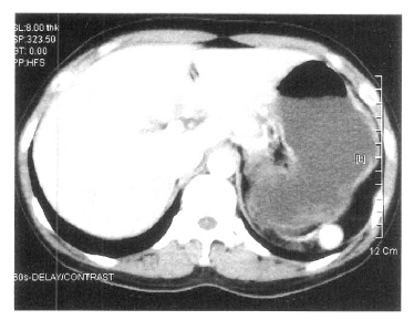
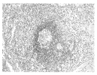
A 37-year-old woman was admitted to our hospital due to gum bleeding and petechiae in both lower extremities for three days. She had a known diagnosis of AITP and had undergone a splenectomy 11 years ago. She denied taking any medications prior to this hospitalization. On admission, her temperature was 36.6┬░C and her blood pressure was 110/70 mmHg. The physical examination was unremarkable except for the oozing of blood in oral cavity and a diffuse petechiae in both lower extremities. A splenectomy scar was present in her abdomen. The WBC was 7,200/mm3, hemoglobin was 12.4 g/dL, and platelet count was 3,000/mm3. The serum electrolytes and liver chemistry were within normal limits. Serum urea nitrogen and creatinine were normal. A routine urine analysis showed the presence of microscopic hematuria. The coagulation profile was all within the normal limits. Daily prednisolone and intravenous immunoglobulin (IVIG) were started. A spleen scan obtained after the intravenous injection of technetium-99m-labeled denatured RBC revealed a focal uptake in the posterior aspect of the left upper quadrant, and these findings are consistent with the presence of an accessory spleen (Figure 1). A computed tomographic (CT) scan of the abdomen revealed a 2├Ś2 cm sized soft tissue lesion on the left sub-diaphragmatic area (Figure 2). On the 22nd hospital day, an accessory splenectomy was performed and the operation proceeded without complication. A dark brown mass was obtained and the pathologic finding was splenic tissue (Figure 3). The postoperatively platelet count soon increased to 71,000/mm3 and the patient was discharged. Two months after the accessory splenectomy, her platelet count dropped to 5,000/mm3. A repeated follow-up spleen scan did not show any remaining accessory spleen. A bone marrow examination showed there was still adequate megakaryocytes with normal hematopoiesis (Figure 4). She is being managed with oral cyclophosphamide with a stable platelet count in the range of 50000/mm3 at present.
DISCUSSION
AITP is an autoimmune disorder in which the platelets opsonized with anti-platelet autoantibodies, are removed prematurely by the reticuloendothelial system; this leads to a reduced peripheral blood platelet count. Although bone marrow megakaryocytes are often increased, a relative marrow failure may play a role in a proportion of patients3). In the adult form there is usually no obvious antecedent illness and most patients have a chronic thrombocytopenia; spontaneous recovery is rather uncommon. The frequency of death from hemorrhage for patients who failed to achieve an adequate platelet count is 5%4). Treatment is generally indicated for the typical patient who presents with bruising or bleeding5). The standard therapy is oral corticosteroids, intravenous immunoglobulin (IVIG) and splenectomy. In the absence of hemorrhage or another medical emergency, treatment is generally initiated with prednisone and around 20ŌĆō30% of patients can expect a long-term response6). Intravenous immunoglobulin is used to treat internal bleeding, when the platelet count remains below 5,000/mm3 despite treatment with corticosteroids or when extensive or progressive purpura is present. Approximately 80 percent of the patients have a response, but a sustained remission is infrequent7). Most adults relapse during or after discontinuation of the prednisolone, at which time a splenectomy is considered. In general, a splenectomy is the treatment of choice in any AITP patient who requires additional medical treatment unless otherwise contraindicated. Approximately two-thirds of adults will have an initial complete response to splenectomy and another 15% will have a stable partial response8). Approximately 15% of responding patients will relapse either soon after splenectomy or, as is less common, many years later5). The presence of an accessory spleen should be sought in any patient who has relapsed and is likely to require additional treatment. Accessory spleens can be located in many sites (from the splenic hilum to the scrotum) and these are sometimes surrounded by fatty tissue that impairs their visualization9). The next approach for patients that are suspected of having residual splenic tissue is to detect its presence with magnetic resonance imaging or with other sensitive scanning techniques. Less than one-quarter of these patients will have a long-term remission after the removal of an accessory spleen, and this is probably due to increased destruction of platelets by accessory parts of the resticuloendothelial system other than the spleen. If there is no response after splenectomy, prednisone is reintroduced or, the therapy is changed to the immunosuppressive drugs danazol or high-dose dexamethasone10). Cyclosporin A has been shown to increase the platelet count when it is given either alone or with prednisolone.
In our patient, removal of the accessory spleen resulted in only a transient increase in the platelet count and she required additional immunosuppressive agents.


 PDF Links
 PubReader
 ePub Link
 Full text via DOI
 Download Citation
 Print
Print


