


INTRODUCTION
Primary gastrointestinal lymphoma occurs in between 1 to 4% of all gastrointestinal malignancies. The gastrointestinal tract is the most common site of extranodal lymphomas, and colorectal lymphoma comprises between 10 to 20% of primary gut lymphomas in a larger published series1). Primary colorectal lymphoma accounts for only about 0.2% of large intestinal malignancies2). Most such lymphomas are B-cell origin, and T-cell lymphoma of the gastrointestinal tract is extremely rare3).
There is a growing interest in the combination of gastrointestinal malignancy and chronic inflammatory bowel disease. Ulcerative colitis is probably best known to be associated with primary lymphoma of the colorectum. Recently, there have been reports suggesting an association of colorectal lymphoma with Crohns disease. Because the clinical symptoms of chronic inflammatory bowel disease and malignant lymphoma of the colon can be very similar, diagnosis of lymphoma can be masked and correct treatment may be delayed4). We encountered a patient with primary T-cell lymphoma of the colon.
CASE REPORT
A 40-year-old woman suffering from abdominal pain and diarrhea since September 1994 was admitted to another institution. She had undergone colonoscopic examination and endoscopy of the colon and histological examination showed chronic inflammatory bowel disease with signs of Crohns disease involving the entire colon and the terminal ileum. Differential therapeutic strategies, including corticosteroid, had improved the symptoms which were dominated by abdominal pain and bloody stools 5 times a day for 6 months. After stopping medication, including corticosteroid since Februrary 1995, she had experienced abdominal pain and bloody stool twice a day.
When she visited our institute in April of 1995 due to ineffective long-term treatment with corticosteroid, she presented with bloody stool twice a day and 7kg weight loss in a period of six months. Among her complaints were having diarrhea over 5 times a day and bloody stool twice a day. She also complained of dizziness, anorexia and general weakness of the body. Physical examination revealed a slightly slender female. Her blood pressure was 130/80mmHg, pulse rate 85/min, respiratory rate 20/min and body temperature 36.┬░C. There was no abnormal sound in the chest. Abdominal palpatation and percussion indicated a slightly painful abdomen and showed normal peristalsis, no superficial lymphadenopathy and no hepatospelomegaly. Bowel sounds were decreased.
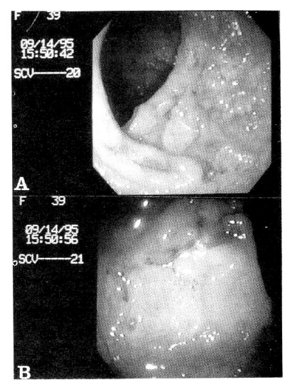
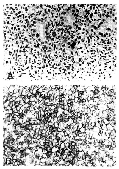
The hematocrit was 38.6% and the white cell count was 7300/mm3, with 89% polymorphonuclear cells and 5% lymphocyte. The serum sodium was 138mEq/L, potassium 4.0mEq/L, chloride 99mEq/L, bicarbonate 23.2mEq/L. The results of liver function tests were as follows : protein 7.3g/dl, albumin 4.4g/dl, cholesterol 189mg/dl, bilirubin 0.6mg/dl, alkaline phosphatase 69U/L, AST 17U/L and ALT 13U/L. Urinalysis did not reveal any evidence of proteinuria. The result of a stool examination revealed positive occult blood. Anti-HIV was negative, findings on the chest radiograph and simple abdomen were normal. Colon study revealed shallow and geographic ulcerations along the entire colon. Small bowel series and enteroclysis revealed ulceration on the distal jejunum. Laboratory examination showed mild inflammatory activity, anemia, normal total and differential white blood cell counts and normal immunoglobulin values. Repeated stool cultures were negative. A chest X-ray showed no evidence of lymphadenopathy. Abdominal ultrasonography and computed tomography were both negative. In upper endoscopy, we were able to find only multiple erosions of the gastric mucosa. Histological examination of the stomach showed only inflammatory infiltrate. Colonoscopy showed geographic ulcerations along the entire colon, especially rectum and terminal ileum(Fig. 1). Histologic examination of the colon showed diffuse infiltration of monotonous small atypical lymphocytes in the laminar propria of the colon(Fig. 2A). These atypical lymphocytes invaded the epithelium of the crypts, mimicking a lymphoepithelial lesion of low grade B cell lymphoma of MALT type. However, they were positive for CD45RO(Fig. 2B) and CD3, negative for CD20. Thus, a diagnosis of primary T-cell lymphoma, small cell type, was made. Although there was reactive epithelial change including depletion, no histologic evidence of inflammtory bowel disease was found in the uninvolved mucosa. Also, we reviewed the pathology of the colon which was taken from colonoscopy at another institute. The histologic findings were very similar to those of the 2nd biopsy except for marked acute inflammatory reaction associated with mucosal ulceration. There were dense atypical small lympohoid infiltrates in the lamina propria with destruction of colonic glands and crypts. No evidence of Crohns disease was present. The histologic finding of bone marrow was negative. In conclusion, she had developed primary T-cell lymphoma of the colon.
DISCUSSION
To distinguish between primary and secondary gastrointestinal lymphoma, the definition of primary gastrointestinal lymphoma5) is quite applicable, which consists of no palpable superficial lymphadenopathy at presentation, a normal chest X-ray, no evidence of hepatosplenomegaly, normal white blood cell count(i.e., no evidence of leukemia) and a predominant tumor mass in the bowel with only local lymphadenopathy. This case fulfills these criteria and we considered it as a primary lymphoma which developed in the colon.
Most primary gastrointestinal lymphoma are of B-cell origin. Only a few cases are reported to be T-cell marker positive3). The most common site of involvement by gastrointestinal T-cell lymphoma is the small bowel and other sites are exceptional. T-cell malignant lymphoma of the gastrointestinal tract, although relatively infrequent, may present a difficult diagnostic challenge to pathologists.
Difficulties in diagnosing T-cell lymphomas of the gastrointestinal tract stem from several sources. Ideally, tissues from these tumors should be frozen for immunostaining of cell surface markers and for gene rearrangement studies, as well as being fixed routinely and embedded in paraffin. Moreover, even when such tissues are frozen, the florid inflammatory infilatrate which usually accompanies these neoplasms, often with ulceration and necrosis, may be all that is present in the areas sampled, or inflammatory cells may greatly outnumber the tumor cells that the neoplasms are difficult to be detected in the frozen section. In this case, colonoscopic and histologic finding showed ulceration and inflammation of the entire colon mimicking Crohns disease. Therefore, primary T-cell lymphoma was not diagnosed during the first examination.
An ideal solution to these problems in diagnosis would be to use antibodies for detection of T-cell in routinely fixed, paraffin-embedded tissue. This would enable appopriate blocks from resection specimens or biopsy specimens to be selected on a basis of well-preserved morphology, and would allow characterization of the neoplastic cells in their inflammatory background. A number of antibodies reactive with routinely processed tissue have proven recently to be effective in detecting B-and T-cell and their malignant counterparts in routinely processed specimens from nodal and extranodal malignant lymphomas6).
Also, there was the combination of gastrointestinal malignancy and chronic inflammatory bowel disease. It is already well known that after 25ŌĆō30 years of ulcerative colitis, the cumulative risk of developing cancer is about 10%7). Adenocarcinoma complicating Crohns disease is increasingly recognized, especially in the small intestine8). The nature of the relation between inflammatory bowel disease and intestinal lymphoma, however, is less certain. The first instance of this association was evaluated by Barken, in 1927, who reported two cases of ulcerative colitis with malignant lymphoma. The small number of reported cases makes it difficult to prove definite association between ulcerative colitis and colonic lymphoma.
Treatment of gastrointestinal lymphoma primarily follows the guidelines for lymphoma treatment. Today, the principle treatment for non-Hodgkins lymphoma is a combination of chemotherapy with radiotherapy. Surgery is used chiefly as a diagnostic tool, with some unique exceptions. Combination chemotherapy with cyclophosphamide, doxorubicin, vincristine and prednisone-CHOP regimen was introduced in the 1970's. Since then, a number of second generation and third generation regimens and combinations of non-cross-resisitant regimens for non-Hodgkins lymphoma have been developed. The benefit of these newer regimens is now in question, but some issues are not yet clearly resolved. Current trials of chemotherapy for non-Hodgkins lymphoma may hold the answers. However, combination chemotherapy with CHOP regimen should be the primary treatment in cases of lymphoma which is widespread throughout the gastrointestinal tract. Radiotherapy, chemotherapy, or both, may also be of use in cases of advanced stage and high grade lymphoma. However, prognosis is poor. Survival of patients with peripheral T-cell lymphoma tends to be short. It is noteworthy that the two reported cases of T-cell lympho¬ma both pursued an aggressive and fatal course9).
The traditional approach of the frozen section immunohistochemistry, so valuable for characterizing most maligant lymphomas, is often less helpful in the evaluation of the typically diffusely involved, inflammated and ulcerated resection specimens. Use of paraffin-reactive monoclonal antibodies facilitated the diagnosis. In conclusion, this is a report of primary T-cell lymphoma of the colon mimicking CrohnŌĆÖs disease.
 PDF Links
 PubReader
 ePub Link
 Full text via DOI
 Download Citation
 Print
Print


