


INTRODUCTION
In 1932, Crohn, Ginzburg and Oppenheimer first presented the clinical and pathologic features of a new disease which affected the terminal ileum. They described the disease as granulomatous inflammation associated with thickening of the bowel wall, and called the condition ŌĆ£terminal ileitisŌĆØ1). It later became apparent, however, that any region of the gastrointestinal fract from the mouth to the anus could be affected, so its name was changed from regional ileitis to regional enteritis or CrohnŌĆÖs disease.
Interest in this disease has increased recently, because its incidence and prevalence have greatly increased in western countries since the beginning of the sixties.
The disease is characterized by its insidious onset with diarrhea, intermittent abdominal pain, general malaise, loss of weight and anemia; by its indolent, prolonged and variable course; by its diversity of clinical features; by its perianal and sytemic complications; and its remarkable tendency to recur after resection of the involved intestine.
CrohnŌĆÖs disease is quite rare in Korea. It is not well understood why the disorder is rare in korea, but diarrhea of uncertain etiology and intestinal tuberculosis are common among koreans, and there is a possibility that the true incidence of CrohnŌĆÖs disease have been masked.
MATERIALS AND METHOD
The subjects were 45 patients with CrohnŌĆÖs disease, including 23 cases at the authorŌĆÖs of the hospital, that had been reported in korea during the period of 34 years from 1852 to 1985. Nearly all of the cases were diagnosed having CrohnŌĆÖ disease after surgical intervention and subsequent pathologic exam of the resected specimens because they had initially appeared to have apparent surgical problems such as colon cancer, intestinal perforation, intestinal obstruction or acute appendicitis.
On a retrospective basis, 45 patients were reviewed2ŌĆō9) and the following details were recorded: sex, age of onset, site of macroscopic disease at diagnosis, the frequency of clinical symptoms and signs, the incidence of perianal disease, the incidence of extraintestinal manifestations, and the list of presumptive diagnosis on initial evaluation. And, we also compared the results of our patient population with representative experiences reported in the medical literatures.
RESULTS
The total number of the patients was only 45, and of the 45 patients, 25 were males. The male to female ratio was 1.3 to 1 with a slight preponderace to male.
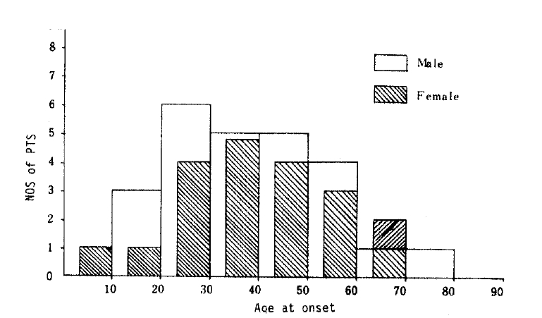
The age at diagnosis (Fig. 1) ranged from 8 to 72 years, and the mean age was 35.5. The disease was relatively uncommon before the age of ten, and the peak incidence occurred in the 3rd, 4th and 5th decades and declined therafter. Nearly one third had the disease diagnosed before their 30th birthday, two thirds between their 20th and 50th birthday, and three fourths before their 50th birthday. One out of ten was more than 60 years of age at diagnosis.
The site of macroscopic disease at diagnosis is shown in Table 1. The patients with CrohnŌĆÖs disease were divided into those with small bowel disease alone, those with ileocolitis, those with disease involving only the colon and the rectum. Nearly 60 per cent of the patients had macrosocpic disease confined to the ileum, and the ileum was involved in more than 75 per cent of the patients. The colon was also involved in one third of the patients, and in only 20 per cent of the cases is the colon affected without concomitant small bowel disease, illustrating the diffuse nature of the disorder. It appears that the vast majority of small and large bowel diseases mainly involve the ileum, cecum and ascending colon. Four out of 45 patients had rectal involvement. Two patients had macroscopic diseases confined to the rectum, two to the sigmoid, and one to the descending colon. One patient had macroscopic involvement of the duodenum.
Table 2 shows representative experience reported in the medical literatures, and of our patient population. The proportion of patients with small bowel, small and large bowel, and large bowel disease at diagnosis shows a significantly greater portion of patients with distal small bowel involvement compared with the patients studied by other authors. There was a corresponding significant reduction in the proportion of patients with macroscopic colonic disease at diagnosis.
Comparison of the anatomic distribution of lesions of CrohnŌĆÖs disease in different centers is shown in Table 3. It is worthy of notice that the anatomic distribution of lesions of CrohnŌĆÖs disease in our patients is quite similar to that observed by Dr. Shapiro 50 years ago. The proportion of the patients with macroscopic disease confined to large bowel alone was only 15.6 per cent.
The frequency of clinical symptoms according to disease location is shown in Table 4. The most common symptom observed was abdominal pain, occurring in 89 per cent of the patients. Diarrhea, however, was observed in only a minority of the patients. Rectal bleeding which was grossly recognizable occurred in 13 per cent of the patients. Nonspecific symptoms such as anorexia, indigestion, weight loss were observed in one fourth of the patients.
Table 5 shows the frequency of clinical signs according to the location of the disease. Abdominal mass was palpable in more than half the cases, which made it difficult to differentiate CrohnŌĆÖs disease from cancer of colon, especially from the one with predominant infiltration of the bowel wall and secondary ulcer formation. The frequency of perianal disease was also low in our patients, occurring in 11 per cent. Formation of internal fistula was found in 9 per cent of the patients.
Extraintestinal findings and their relationship to disease sites are shown in Table 7. In our series of 45 patients, only three patients presented with symptoms of arthritis. Two of them had the disease confined to the small and large bowels. Other extraintestinal manifestations such as skin lesions, hepatobiliary manifestations, renal complications, eye complications were not observed at all.
Table 8 shows the list of diagnosis which had been originally made on initial evaluation. Only 7 out of 45 patients had been presumed to have CrohnŌĆÖs disease from the onset. Two fifths of the patients had been diagnosed colon cancer, because abdominal mass had been palpable in the majority of these patients. Ten patients had been initially thought to have intestinal tuberculosis, and two patients who had been presumed to have acute appendicitis turned out to have CrohnŌĆÖs disease after surgical intervention.
DISCUSSION
It is now known that there are highly varying difference in the reported incidence of inflammatory bowel disease throughout the world and there are no areas which careful search has found to be free of these disorders10).
Hahn3) and Choi4) in our hospital reported 23 cases of CrohnŌĆÖs disease. A review of the literature shows another 22 cases were reported in Korea by 19855ŌĆō9). So the total number of the patients with CrohnŌĆÖs disease that had been reported in Korea during the period of 34 years from 1952 to 1985 only 45. It is not well understood why the disorder is so rare in Korea, but there is a possibility that the true incidence of CrohnŌĆÖs disease would have been masked because of confusion with intestinal tuberculosis and diarrhea of uncertain etiology which are quite common in Korea.
Table 9 is a list of illnesses that could potentially be considered in the differential diagnosis of CrohnŌĆÖs disease in Korea. When, as is often the case, the disease begins abruptly with an apparantly transient episode of diarrhea, abdominal cramps and fever, the physician is likely to pass this initial episode off as salmonella infection which is quite endemic in Korea11). During the early, acute phase of the illness, CrohnŌĆÖs disease may also be confused with appendicitis11). Tuberculous involvement of bowel and peritoneum remains a major medical problem in Korea, and it is extremely difficult to differentiate intestinal tuberoulosis from CrohnŌĆÖs disease because the two disease share cardinal features, including diarrhea, abdominal pain, fever, weight loss, and radiologic demonstration of ileocecal involvement, with X-ray findings of intramural swelling, stricture and fistulas. Amebiasis, which is still common in Korea, may be difficult to differentiate from CrohnŌĆÖ s disease, especially when localized to terminal ileum and cecum causing a chronic intramural lesion11). Because the abdominal mass is palpable in more than half the cases, it is difficult to make a differential diagnosis of CrohnŌĆÖs disease from colon cancer, particularly if the latter lesion predominantly infiltrates the bowel wall and associated with secondary ulcer formation, which sometimes causes sinus tracts and fistulas into bowel or bladder11).
The ratio of male to female patients was 1.3:1 (25 males and 20 females). The difference from those observed by others was not significant12,13). The age at diagnosis of the patients did not show a typical bimodal distribution which had been observed in other series12,13).
A review of the literature shows that there has been considerable change in the anatomic distribution of CrohnŌĆÖs disease over the past 50 years, and the proportion of patients with small bowel, small and large bowel, and large bowel disease at diagnosis shows recently a significantly greater proportion of patients with large bowel involvement at diagnosis13). There was a corresponding significant reduction in the proportion of patients with macroscopic ileal disease at diagnosis. This change has been designated ŌĆ£colonic shiftŌĆØ by some authors14,15). It is quite interesting to find the anatomic distribution of lesions of CrohnŌĆÖs disease in our patients is similar to that observed by Dr. Shapiro 50 years ago (Table 3).
The literature shows that the symptoms of diarrhea is one of cardinal features of CrohnŌĆÖs disease and is present in virtually all patients16). But, in our patients group, diarrhea was observed in only 13 per cent of the patients. The exact reason why the symptom was rarely observed in the CrohnŌĆÖs disease in Korea is not well understood, but there is a possibility that CrohnŌĆÖs disease presenting diarrhea as its chief clinical manifestation has been overlooked, because diarrhea of uncertain etiology is still prevalent in Korea, masking the true incidence of CrohnŌĆÖs disease.
An abdominal mass was palpable in more than half the cases, which made it difficult to differentiate CrohnŌĆÖs disease from colon cancers. Over 40 per cent of the patients had been presumed to have colon cancer on initial evaluation, and that is one of the reasons why most cases in Korea have been reported by surgeons.
Intestinal obstruction was observed in 11 per cent of the patients, and the frequency of the lesion was quite lower than those reported by other authors16).
The frequency of occurrence and detection of perianal disease is known to vary with their classification and the care of the examination, and it has also been shown that perianal disease is more common in CrohnŌĆÖs disease and one half with CrohnŌĆÖs colitis develop symptomatic perianal or perirectal fistulas11,17).
The overall incidence of perianal disease in our patients was only 11 per cent, and it is suggested that low incidence of perianal disease might be due to small percentage of the disases with macroscopic involvement of large bowel.
It is now known that systemic manifestations develop in CrohnŌĆÖs disease, particularly in those individuals who have colonic involvement, about as often as they occur in ulcerative colitis16ŌĆō19). Extraintestinal manifestations also appear to occur more frequently among patients with perianal disease17). In our series of 45 patients, only three patients presented symptoms of arthritis, and other extraintestinal manifestations such as skin lesions, hepatobiliary manifestations, renal complications, eye complications were not observed at all.
In conclusion, CrohnŌĆÖs disease is a quite rare disease among Koreans, so it may be that this review on inflammatory bowel disease cannot be given in statistically meaningful terms, and it would be better to accept the results of the data as a simple indication of the existence of CrohnŌĆÖs disease among Korean people.
 PDF Links
 PubReader
 ePub Link
 Full text via DOI
 Download Citation
 Print
Print


