


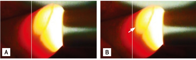
A 62-year-old man, without a medical history of cardiovascular disease, was transferred to our hospital due to sudden-onset chest pain. His blood pressure was 101/21 mmHg, and heart rate was 87 beats per minute. A physical examination revealed a diastolic murmur, but we could not find an obvious Quincke pulse, only by nail bed pressure. However, applying Quincke's pocket f lashlight method revealed momentary accentuation of reddening of the nail bed and fingertips of the left hand (Fig. 1 and Supplementary Video 1 [available online at http://www.kjim.org/]). We suspected aortic regurgitation associated with an aortic dissection, which was conf irmed by trans-thoracic echocardiography and contrast-enhanced computed tomography. Therefore, we performed an emergency operation, and his clinical course was good.
Aortic regurgitation is one of the most common causes of death in patients with an aortic dissection. However, the sensitivity of auscultation for detecting a diastolic murmur is very low. Therefore, at our institution, we have added several other physical signs and imaging modalities to diagnose aortic regurgitation in clinical practice. The Quincke pulse is a famous physical sign, but it is also difficult to define without a microscope and microcapillary tonometer. To address this problem, introduced the pocket f lashlight method, whereby a light source is used to illuminate the fingers. We diagnosed aortic regurgitation based on a diastolic murmur and the Quincke pulse as determined using the pocket flashlight method. This method is a simple tool for diagnosing aortic regurgitation in patients with chest pain.
 PDF Links
 PubReader
 ePub Link
 Full text via DOI
 Download Citation
 Print
Print


