


To the Editor,
Congenital coronary artery anomalies are infrequently seen during coronary angiographic study, reportedly occurring in 0.64% to 1.3% of patients [1]. Most patients with a congenital coronary artery anomaly are asymptomatic; in rare cases, they may present with chest pain and have myocardial ischemia or other life-threatening conditions. We present a patient with the right coronary artery (RCA) originating from the terminal branch of the left circumflex (LCx) artery who presented with chest pain and was found to have a moderate stenotic lesion in the distal LCx artery. Fractional flow reserve (FFR) allowed us to evaluate this moderate stenotic lesion in a single coronary artery, and to treat with medical therapy only.
A 63-year-old man was admitted to the cardiology unit with atypical chest pain. He had hypertension as coronary heart disease risk factor. His vital signs were stable, and physical examination showed no abnormalities. A resting electrocardiogram demonstrated a convex-shaped ST-segment elevation in the precordial lead without reciprocal change. Laboratory findings including cardiac markers showed no abnormalities. Echocardiography revealed normal left ventricular morphology, no regional wall motion abnormality, and a diastolic dysfunction of relaxation abnormality. The patient underwent coronary computed tomographic (CT) angiography. It demonstrated the presence of a single coronary artery with the LCx artery continuing in the course of the RCA and minimal possibility of chronic total occlusion (CTO) of the RCA ostium with a collateral supply from the LCx artery (Fig. 1). It also showed 60% diameter stenosis in the distal LCx artery (Fig. 1).
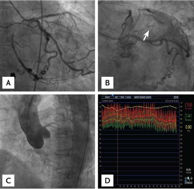
To differentiate single coronary artery from CTO of the RCA ostium and confirm the degree of stenosis in the distal LCx artery, we performed coronary angiography. Selective left coronary angiography displayed normal origin and course of the left main, LCx, and left anterior descending arteries. The LCx artery did not terminate after reaching the crux; giving rise to the posterior descending branch, it coursed in the right atrioventricular groove as if it were the RCA and ended when it reached the right sinus of Valsalva (Fig. 2A). There was a stenosis of 60% diameter in the distal part of the LCx proper where the RCA arose (Fig. 2B). Attempts to engage the right coronary catheter into the RCA ostium were futile, and aortography obtained in the left anterior-oblique projection displayed the absence of the ostium of the RCA in the right sinus of Valsalva (Fig. 2C). To decide the functional significance of the stenosis in the distal LCx artery, we performed an FFR study with a pressure wire and found that there was no critical functional stenosis (FFR, 0.9) (Fig. 2D). The patient was discharged with medications of aspirin, statin, nitrate, and β blocker and free from chest pain.
A single coronary artery is an extremely rare congenital anomaly characterized by a single coronary artery ostium from an aortic sinus, and is seen in only 0.024% to 0.066% of patients who undergo conventional coronary angiography [1]. A single coronary artery is usually asymptomatic and has a benign course, and these patients have a normal life expectancy. The presence of a single coronary artery may bring about myocardial ischemia because of the inability to adequately sustain normal coronary circulation [2].
FFR is a physiologic parameter that can be readily measured during the invasive procedure and can evaluate the functional significance of coronary stenosis. In particular, FFR plays an important role when making a decision for percutaneous coronary intervention (PCI) of angiographically moderate stenosis and assessing accurately the functional consequences of a given coronary stenosis with unclear hemodynamic significance [3]. In addition, the clinical outcome of patients in whom PCI is deferred because FFR indicated no hemodynamically significant stenosis was very favorable [4]. In this case, FFR was a good assessment modality because the lesion of interest in the LCx artery was a part of a single coronary artery that supplied both the LCx artery and RCA territories. With FFR, we could make a decision for deferred PCI and choose medical treatment, even though coronary CT angiography and coronary angiography showed moderate stenosis in the LCx artery.
In conclusion, FFR could successfully evaluate the functional significance of a moderate stenotic lesion in a single coronary artery where the RCA originated from the terminal portion of the LCx artery.

 PDF Links
 PubReader
 ePub Link
 Full text via DOI
 Download Citation
 Print
Print


