


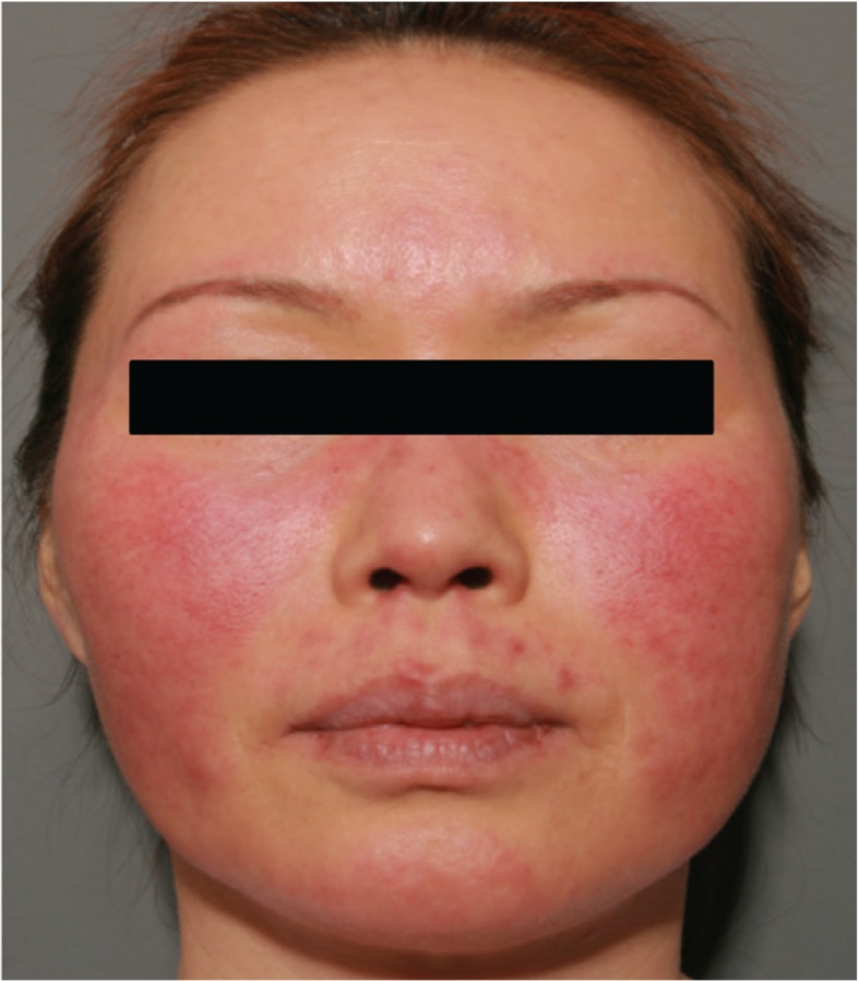
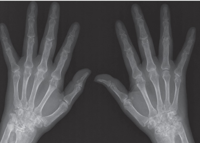
A 45-year-old female presented with a facial rash for 2 weeks. She also had arthralgia of the hand and wrist joints with morning stiffness persisting for 1 hour. On physical examination, there was a typical malar rash on her face (Fig. 1). She had a swan-neck deformity of her left third finger and swelling and tenderness of the bilateral metacarpophalangeal (MCP), proximal interphalangeal (PIP), wrist, and knee joints. The laboratory results were as follows: erythrocyte sedimentation rate 91 mm/hr (normal, < 20), C-reactive protein 2.69 mg/dL (normal, 0.01 to 0.3), white blood cell 3,110/┬ĄL (normal, 4,000 to 10,000, lymphocytes 27.7%), hemoglobin 9.6 g/dL, platelet 190,000/┬ĄL, anti-nuclear antibody 1:40 (a mixed homogenous and speckled pattern), anti-dsDNA antibody 14.2 IU/mL (normal, < 7.0), rheumatoid factor 98 IU/mL (normal, 3 to 18), and anti-CCP antibody > 300 U/mL (normal, < 5). In addition, the serum complement levels were decreased with C3 62 mg/dL (normal, 86 to 160), and C4 6.20 mg/dL (normal, 17 to 47), and the direct Coombs' test was positive for immunoglobulin G. The urine protein/creatinine ratio was 561.93 mg/g. However, anti-cardiolipin, anti-╬▓2 glycoprotein 1, and lupus anticoagulant antibodies were negative. Plain radiographs of both hands showed periarticular osteopenia, joint space narrowing, and marginal erosions at the MCP, PIP, and carpal joints (Fig. 2). Based on the clinical, laboratory, and radiographic results, she was diagnosed with both rheumatoid arthritis and systemic lupus erythematosus or "rhupus syndrome." She was treated with hydroxychloroquine 400 mg, celecoxib 200 mg, prednisolone 5 mg, and methotrexate 10 mg/week and her arthralgia and facial malar rash improved.
 PDF Links
 PubReader
 ePub Link
 Full text via DOI
 Download Citation
 Print
Print


 |
 |