


To the Editor,
Atrial septal defect (ASD) is one of the most common types of congenital heart disease in adults [1]. Surgical closure has been accepted as the standard treatment for clinically significant ASD; however, in ostium secundum ASD, catheter-based closure could be an alternative option. The first case of transcatheter ASD repair was reported in 1974 by King et al. [2]. Since then, remarkable advances in the transcatheter closure technique and revolutionary changes in device technology have occurred. Therefore, transcatheter closure is now considered a primary treatment option for ostium secundum ASD patients [1,3]. In contrast, transcatheter closure for other types of ASD is not recommended [1]. For such patients, surgical repair is the standard treatment option [4].
A previous report described a woman with inferior sinus venosus ASD who received septal defect repair using the transcatheter technique; however, the closure procedure was unsuccessful and she subsequently underwent surgical repair [5]. Here, we describe a case of successful transcatheter closure for an inferior sinus venosus ASD.
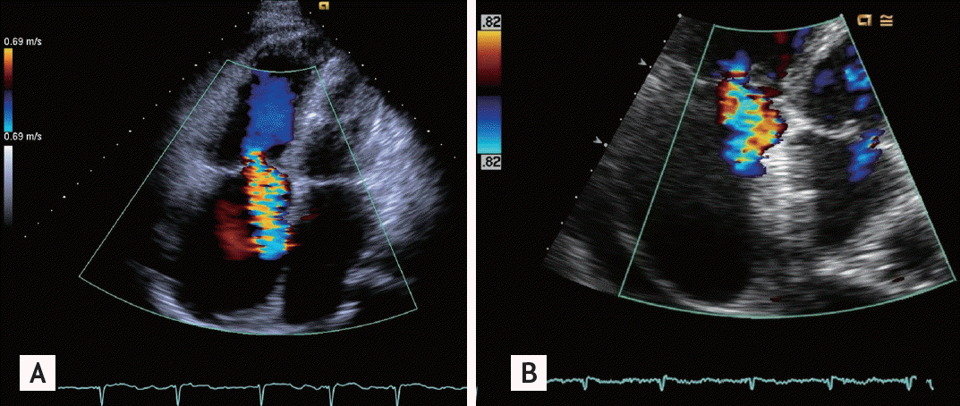
A 76-year-old female presented with New York Heart Association class IV dyspnea. She had a history of hypertension and atrial fibrillation and was taking diuretics, angiotensin receptor blockers, amiodarone, and warfarin. On physical examination, an irregular heartbeat with a pansystolic murmur was auscultated, and pitting edema was observed. Initial arterial blood gas analysis results showed hypoxemia. Cardiomegaly was noted on a chest radiograph. Echocardiography revealed dilation of the right atrium, right ventricle, and left atrium, as well as severe tricuspid regurgitation (Fig. 1A) with pulmonary hypertension (the right ventricular systolic pressure was 75 mmHg and the tricuspid valve showed poor coaptation with a D-shaped left ventricle; the tricuspid annular plane systolic excursion [TAPSE] was 1.6 cm). Doppler echocardiography showed a turbulent shunt flow near the inferior vena cava (IVC).
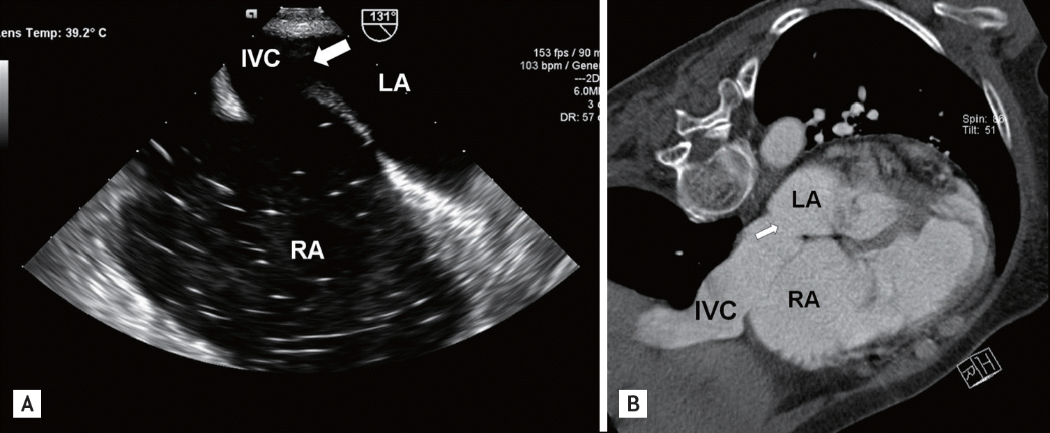
To clarify the anatomical features of the shunt, transesophageal echocardiography was performed. The results showed an inter-ASD near the IVC with a size of approximately 1.74 cm (Fig. 2A). Chest computed tomography revealed an approximately 1.7 cm-sized inferior venosus ASD (Fig. 2B).
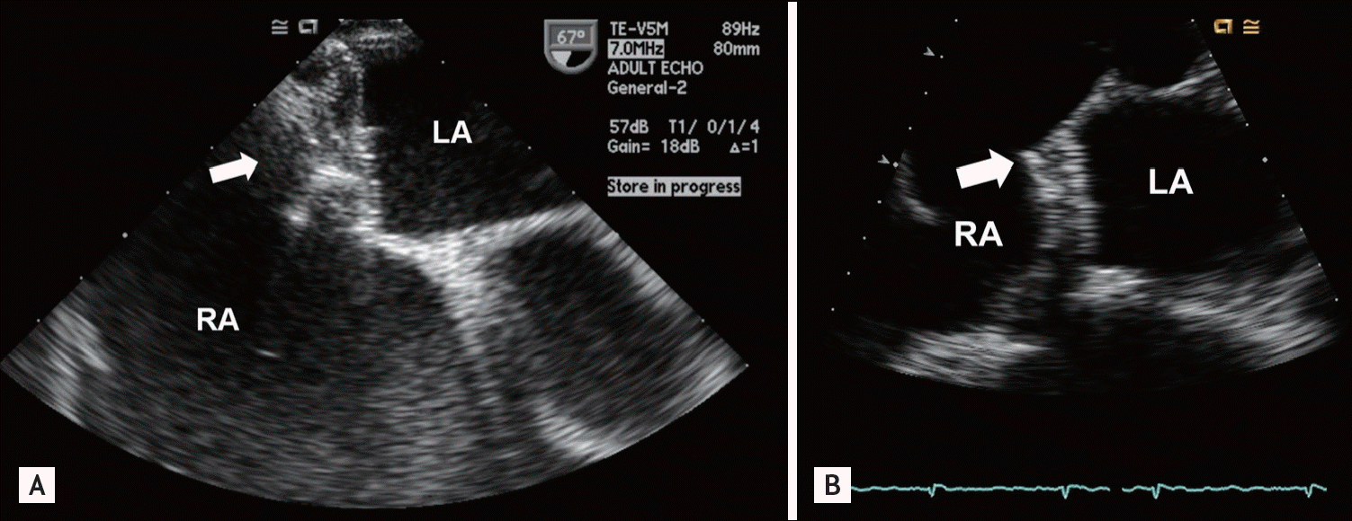
Although elective surgical repair of the defect is normally the treatment of choice, the patientŌĆÖs general condition and her age were not suitable for general anesthesia, and she did not want to receive surgery. Typical inferior sinus venosus ASD is a contraindication for transcatheter closure, but we decided to repair the septal defect using the transcatheter technique because of the atypical features of the septal defect; it was located away from the tricuspid valve, and there was a small ridge on the posterior side of the defect. Using the transcatheter approach, a 20 mm-sized Amplatzer occluder device (St. Jude Medical, St. Paul, MN, USA) was successfully applied in the defect. There were no acute complications after the procedure, and the surgery was well tolerated. After the closure, the patientŌĆÖs dyspnea improved dramatically and her hypoxemia recovered. On follow-up echocardiography, there was no abnormal shunt flow, the occlusion device was well placed (Fig. 3A), and the tricuspid regurgitation (Fig. 1B) and pulmonary hypertension were improved (the right ventricular systolic pressure was 41 mmHg, moderate tricuspid regurgitation was noted, and the TAPSE was 1.92 cm).
A follow-up exam 1 year after surgery showed that the patient was in good condition and did not suffer from dyspnea when performing daily activities. Six months following the procedure, warfarin was switched to dabigatran because of the patientŌĆÖs non-compliance and difficulty achieving the target international normalized ratio; the patient showed good compliance with dabigatran. Follow-up echocardiography 9 months after the procedure showed that the occluder device was well placed, without abnormal shunt flow (Fig. 3B).
Surgical repair is the standard therapeutic option for ASD patients of all ages. However, transcatheter closure has recently received increased acceptance in ASD patients [1,3]. Transcatheter closure guarantees fewer complications and a shorter hospital stay than surgical repair, and both procedures have similar success rates. However, transcatheter closure is indicated only in ostium secundum ASD patients because of its anatomical features.
In this case report, we described the catheter-based repair of an inferior sinus venosus ASD. To the best of our knowledge, this is the first successful case of transcatheter sinus venosus ASD closure. Typical sinus venosus ASD remains a contraindication for transcatheter closure of the defect. However, this case suggests that if a sinus venosus ASD patient cannot undergo surgery due to several causes (e.g., combined comorbidities, general condition, and old age) and the patientŌĆÖs septal defect has anatomical features suitable for device closure (e.g., a rim or small ridge toward the caval vein), transcatheter closure might be an alternative therapeutic option. While it may not be generally applied, transcatheter closure could be considered a secondary option for carefully selected patients.
 PDF Links
 PubReader
 ePub Link
 Full text via DOI
 Download Citation
 Print
Print


