


INTRODUCTION
Familial Mediterranean fever (FMF) is an autosomal recessive disorder characterized by attacks of fever and diffuse abdominal pain. Patients are usually symptom free between the episodes and the frequency of attacks differ from patient to patient [1]. Since FMF usually affects young individuals, a practical challenge emerges when a patient who was not diagnosed with FMF presents classical symptoms of abdominal pain, fever, nausea, and vomiting. The primary concern in this setting is to distinguish FMF attack from appendicitis within clinically critical time interval. In case of perforated appendicitis a surgical emergency might become a catastrophic clinical issue due to the delay in diagnosis considering the vice versa situation unless the physician proceeds with a prompt but not prudent decision to operate the patient considering acute appendicitis, will lead to an unnecessary surgery in an otherwise nonsurgical patient facing the risks of operation [2,3]. Thus, reliable tools are of crucial importance in determining whether the case presentation is due to FMF attack or acute appendicitis.
Several clinical evidence and biochemical markers have introduced to differentiate the two cases safely and efficiently up to date [1-4]; however, no single laboratory or clinical factor derived concrete differentiative outcomes in this regard. Recent clinical evidence proved the association of neutrophil lymphocyte ratio (NLR) with a number of inflammatory markers. In addition, NLR was shown to be associated with adverse clinical outcomes in various clinical settings [5-10]. Previous studies denoted that FMF attack is associated with an increased value of NLR compared with patients without an active attack [11]. A majority of research revealed that NLR values were significantly different between patients with FMF and healthy subjects showing a positive correlation with NLR subclinical inflammation of attack-free individuals [12]. Previous studies have also emphasized that NLR could be a more sensitive predictor of acute appendicitis compared to C-reactive protein (CRP) and total white blood cell (WBC) count [5,6]. Apart from other causes of lower abdominal quadrant pain, acute FMF attack also presents elevations in acute phase markers including WBC count [7], and solely considering the CRP and WBC count may not be sufficient to differentiate FMF attack and acute appendicitis. To the best of our knowledge, there is no study in the literature analyzing the predictive value of NLR in this regard. Thus, we aimed to evaluate inflammatory markers along with NLR in patients with histologically proven appendicitis and FMF attacks in patients that were already diagnosed with FMF.
METHODS
This was a retrospective study to compare NLR along with other laboratory parameters between patients who underwent laparotomy for acute appendicitis and FMF attack with pathological evidence. Twenty-three patients with acute appendicitis and 88 patients with acute FMF attack who applied to the emergency services of two hospitals were enrolled in a retrospective manner. All the FMF patients had previously been diagnosed and were administered regular colchicine treatment (none of these attacks were classified as a surgical or another medical condition other than FMF). Patients with atypical attacks and acute abdominal pain due to causes other than FMF attack were excluded from the study. All acute appendicitis cases with suggestive features were practiced laparotomy that had uncomplicated appendicitis morphologically and pathologically. Patients who underwent laparotomy but appeared to have normal appendices were excluded from the study. The patients with diabetes mellitus, coronary heart diseases, thyroidal dysfunction, anemia, B12 and folic acid deficiency, acute/chronic infection, autoimmune disorders, chronic obstructive pulmonary disease, established malignancy, and history of smoking were excluded. Blood counts and CRP values at presentation were obtained from hospitals’ electronic database systems. A special effort has been exerted to maintain the blood count and CRP values at the outset of clinical presentation and before surgical intervention in acute appendicitis patients. Institutional Review Board of Necmettin Erbakan University granted approval for the study protocol.
Statistical analysis
The statistical evaluation was conducted using the SPSS version 16 (SPSS Inc., Chicago, IL, USA). The Kolmogorov-Smirnov test was applied to check the distribution of parameters. Categorical data were presented as numbers and percentages, and continuous data were expressed as mean ± standard deviation. The difference between the categorical variables was determined using the chi-square test. The independent Student t test was used to determine the difference in normally distributed data, and the Mann-Whitney U test was used for comparison of median for the non-parametrically distributed variables. A p < 0.05 was considered to be statistically significant.
RESULTS
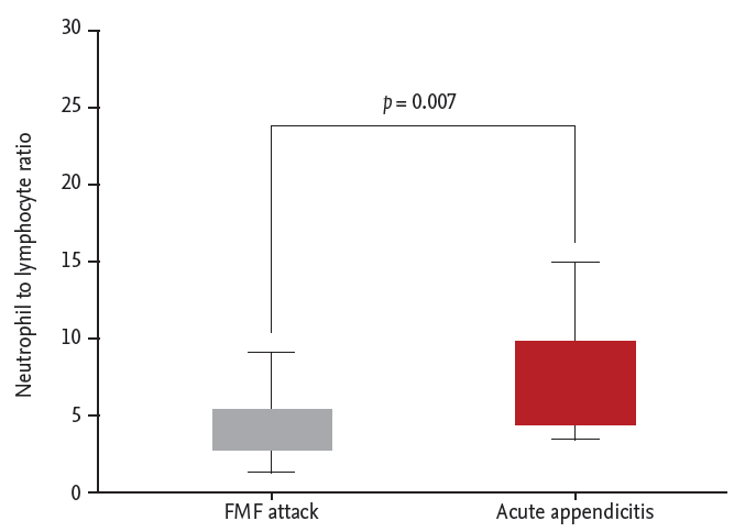
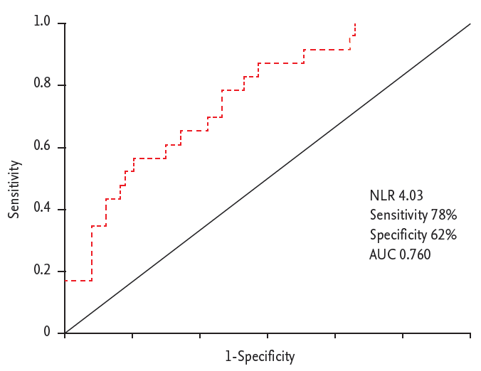
Eighty-eight patients with acute FMF attack (55 females, 33 males; mean age, 31.5 ± 10.4 years) and 23 patients with acute appendicitis (9 females, 14 males; mean age, 30.8 ± 11.9 years) were included in the study. The mean age, gender distribution, CRP and hematologic counts of the groups are depicted in Table 1. NLR was significantly higher in patients with acute appendicitis compared with patients presented with an FMF attack (8.24 ± 6.31 vs. 4.16 ± 2.44, p = 0.007) (Fig. 1). On the other hand, there was not a significant difference between the groups in terms of serum CRP values (39.0 ± 28.1 vs. 28.9 ± 23.3, p = 0.088). Clinical characteristics and physical examination findings of the patients with FMF attack and acute appendicitis are shown in Table 2. We evaluated the performance of NLR in patients with acute appendicitis with receiver operating characteristic analysis, when cut-off is taken as 4.03, its sensitivity, specificity, and area under the curve were found to be as 78%, 62%, and 0.760, respectively (95% confidence interval, 0.655 to 0.865; p < 0.001) (Fig. 2).
DISCUSSION
The prominent finding of this study was that NLR was significantly higher in patients with acute appendicitis compared with patients suffering from an acute FMF attack. To best of our knowledge, this is the first study which utilizes NLR with intend to differ acute appendicitis from an FMF attack.
Presentations of acute appendicitis and FMF attack may have confusing signs and symptoms in some patients especially when the patient with abdominal pain has no previous diagnosis of FMF. Even in patients previously with diagnosed FMF, acute appendicitis may develop and the patient and the caregiver’s attribute can direct to a new FMF attack rather than acute appendicitis. In this case, delay in the diagnosis may lead to increased morbidity an even mortality. In areas where FMF is endemic such as Turkey, many FMF patients had undergone unnecessary abdominal exploration for suspected acute appendicitis at some point during the course of their disease [8]. In one study, the frequency of appendectomy found in FMF patients was far above the reported rate in the general population (40% vs. 12% to 25%) [9]. Previous research has also shown that a significant number of patients who presented with acute abdominal pain and other suggestive features for acute appendicitis have been found to have no histologic features of appendicitis when underwent surgery [10]. Therefore, it is a great clinical challenge to accurately and reliably differ acute appendicitis from an FMF attack.
NLR is a novel inflammatory marker and the value of NLR comes from its readily available nature. Complete blood count is almost an inevitable part of modern laboratory evaluation in many clinical settings and NLR can easily be calculated from the blood count. A number of studies showed that NLR was closely correlated with other markers of inflammation. Moreover, in patients with various cancers [13-15], liver cirrhosis [16], chronic kidney disease [17], and cardiac disease [18,19], NLR could predict adverse clinical outcomes independent of major established risk factors.
Several biochemical parameters have been investigated whether they can reliably differentiate acute appendicitis and acute FMF attack [20]. Serum resistin and visfatin levels have failed to distinguish acute appendicitis from an acute FMF attack [2]. Procalcitonin was found to be significantly higher in patients with appendicitis compared with those of patients with acute FMF attack [4]. However, routine implementation of procalcitonin requires further studies and prohibitively costly yet.
NLR is a simple, non-invasive and cost-effective marker of inflammation in various diseases and is calculated using data obtained from the complete blood count. NLR has been tested to date regarding its ability to accurately diagnose acute appendicitis preoperatively in unselected patients. Almost 20 years ago before NLR became so popular, Goodman et al. [5] declared that 88% of patients with histologically proven appendicitis had a NLR > 3.5. The authors also concluded that NLR was more sensitive to detect acute appendicitis compared with total WBC count. In a retrospective study in children Yazici et al. [21] found that 90.2% of the appendicitis group and 12.3% of the nonspecific abdominal pain group had NLR higher than 3.5. Markar et al. [6] evaluated 1,117 cases who underwent appendectomy in which NLR appears to be of greater diagnostic accuracy than either total WBC count or CRP alone. Moreover, some studies reported that NLR could also predict severity of the appendicitis as Ishizuka et al. [22] showed that NLR > 8 shows a significant association with gangrenous appendicitis in patients undergoing appendectomy. However, none of these studies specifically examined the differential power of NLR between acute appendicitis and FMF attack.
Our results showed that NLR values were significantly higher in patients with acute appendicitis compared with those of acute FMF attack while there was not a significant difference between the groups in terms of serum CRP values. We think that this finding may be particularly important when CRP and clinical findings are not sufficient to safely distinguish acute appendicitis from an FMF attack.
Several limitations of this study are noteworthy. This was a retrospective study and prospective studies recruiting naive patients that admitted emergency services with abdominal pain and/or fever along with other suggestive features of acute appendicitis and/or FMF attack are needed. The differential diagnosis between the two should ideally be based on clinical features at the time of admission and possibly a panel of biochemical tests revealing the degree of inflammation and blood counts. None of the single clinical or laboratory parameters are sensitive or specific enough for concrete diagnosis or exclusion of acute appendicitis or FMF attack. Thus, value of single parameters, in this study example, NLR, should be considered as adjunct to other clinical and laboratory parameters. Prospective studies with large sample size should be designed to achieve a cut-off value or prediction rule based on several clinical and laboratory parameters are definitely needed. Since all FMF patients in our study had their attacks sometime after having diagnosed and received colchicine treatment, these attacks may not be as severe as their first FMF episode which may have caused a more challenging clinical picture. Thus, a prospective study recruiting naive FMF patients would be more ideal to evaluate the role of NLR as a distinction factor. Despite aforementioned limitations, this is the first study showing promising role of NLR at differentiating FMF attack and acute appendicitis.
In conclusion, the main finding of this study can be elaborated as NLR; the simple and cost-effective marker, was significantly higher in patients with acute appendicitis compared with in patients with an FMF attack. We conclude that evaluation of the NLR may have a value in prevention of an unnecessary surgical operation in a non-surgical patient with attendant risks of the operation. Prospective studies are needed to clarify the role of NLR in this situation.
KEY MESSAGE
1. Acute appendicitis and familial Mediterranean fever (FMF) attack may be confused in emergency department and reliable markers to differentiate the two are required.
2. In this study, neutrophil to lymphocyte ratio was significantly higher in patients with acute appendicitis compared with those of FMF attack.
3. Prospective studies taking clinical presentation as well as several laboratory markers into account would provide additional clues to distinguish the two disorders.
 PDF Links
 PubReader
 ePub Link
 Full text via DOI
 Download Citation
 Print
Print


