


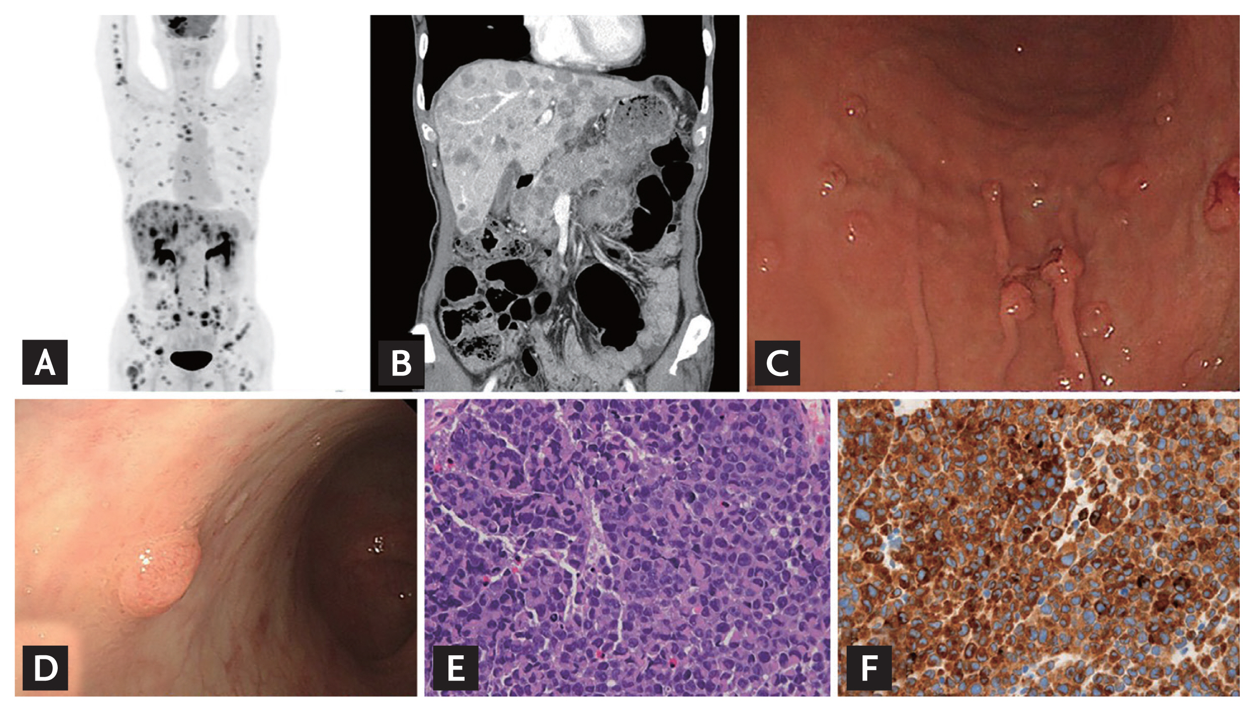
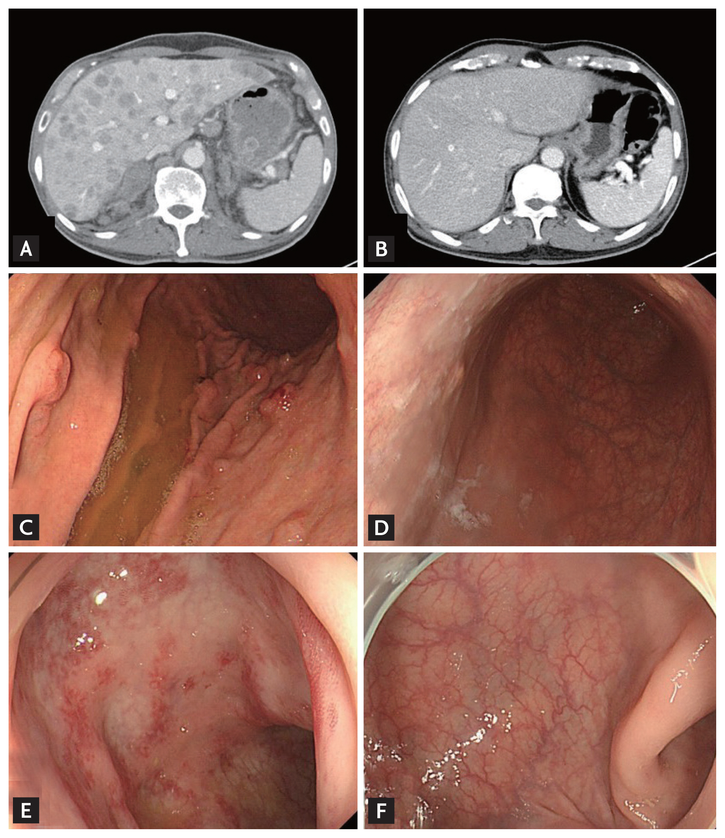
A 64-year-old man presented with multiple metastatic malignant masses throughout the body. (Fig. 1A and 1B). Endoscopic examination revealed umbilicated nodules of various sizes in the stomach (Fig. 1C) and a nodule with peripheral pigmentation in the rectum (Fig. 1D). Malignant melanoma was diagnosed in all endoscopic forceps biopsies (Fig. 1E and 1F). He was diagnosed with malignant melanoma with multiple metastases and an unclear primary site. Nivolumab treatment (human programmed death receptor-1 [PD-1] blocking antibody) was started at 3 mg/kg every 2 weeks. Most metastases and endoscopic malignant nodules disappeared after 6 months of treatment (Fig. 2).
Malignant melanoma can metastasize to all parts of the body and the metastatic rate of the gastrointestinal tract is reportedly around 60%. Initially, metastatic malignant melanoma did not respond to chemotherapy and was considered incurable. However, the advent of immunotherapy dramatically impacted the treatment response of malignant metastatic melanoma. We present comparative pictures showing complete endoscopic regression after PD-1 inhibitor treatment. The patient provided informed consent for publication of this case.
 PDF Links
 PubReader
 ePub Link
 Full text via DOI
 Download Citation
 Print
Print


