


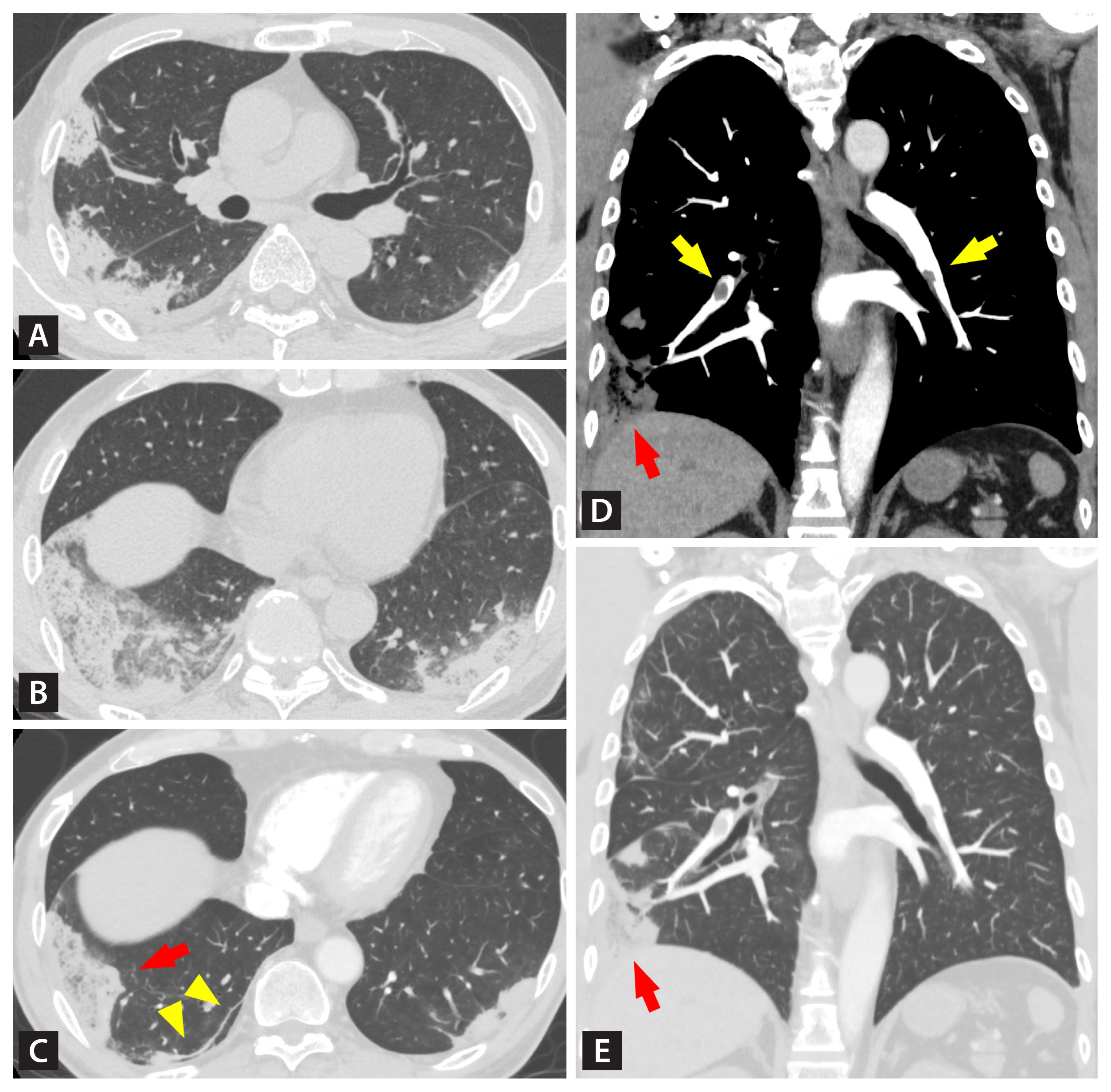
A previously healthy man in his 50s was admitted to our hospital with fever and hemoptysis in late 2020. He had developed fever 17 days before and was subsequently diagnosed with coronavirus disease 2019 (COVID-19). On admission, his oxygen saturation was 96% on room air and D-dimer level was 5,410 ng/mL. Unenhanced chest computed tomography (CT) showed peripheral consolidations with ground-glass opacities and pleural effusion (Fig. 1A and 1B). He was treated with methylprednisolone, favipiravir, and edoxaban.
Hemoptysis persisted and contrast-enhanced CT was performed on day 5. Most consolidations were resolving with a residual subpleural curvilinear line. A wedge-shaped consolidation with internal lucencies persisted suggesting pulmonary infarction (Fig. 1C). Coronal images revealed bilateral pulmonary emboli, one of which corresponded to the remaining consolidation (Fig. 1D and 1E). The pulmonary infiltrates disappeared after 3 months of edoxaban therapy.
Pulmonary embolism is common in patients with COVID-19, while hemoptysis and pulmonary infarction are infrequent. Physicians should perform contrast-enhanced CT for COVID-19 patients with hemoptysis and can continue anticoagulation for pulmonary infarction causing hemoptysis.
Written informed consent was obtained from the patient for publication.
 PDF Links
 PubReader
 ePub Link
 Full text via DOI
 Download Citation
 Print
Print


