


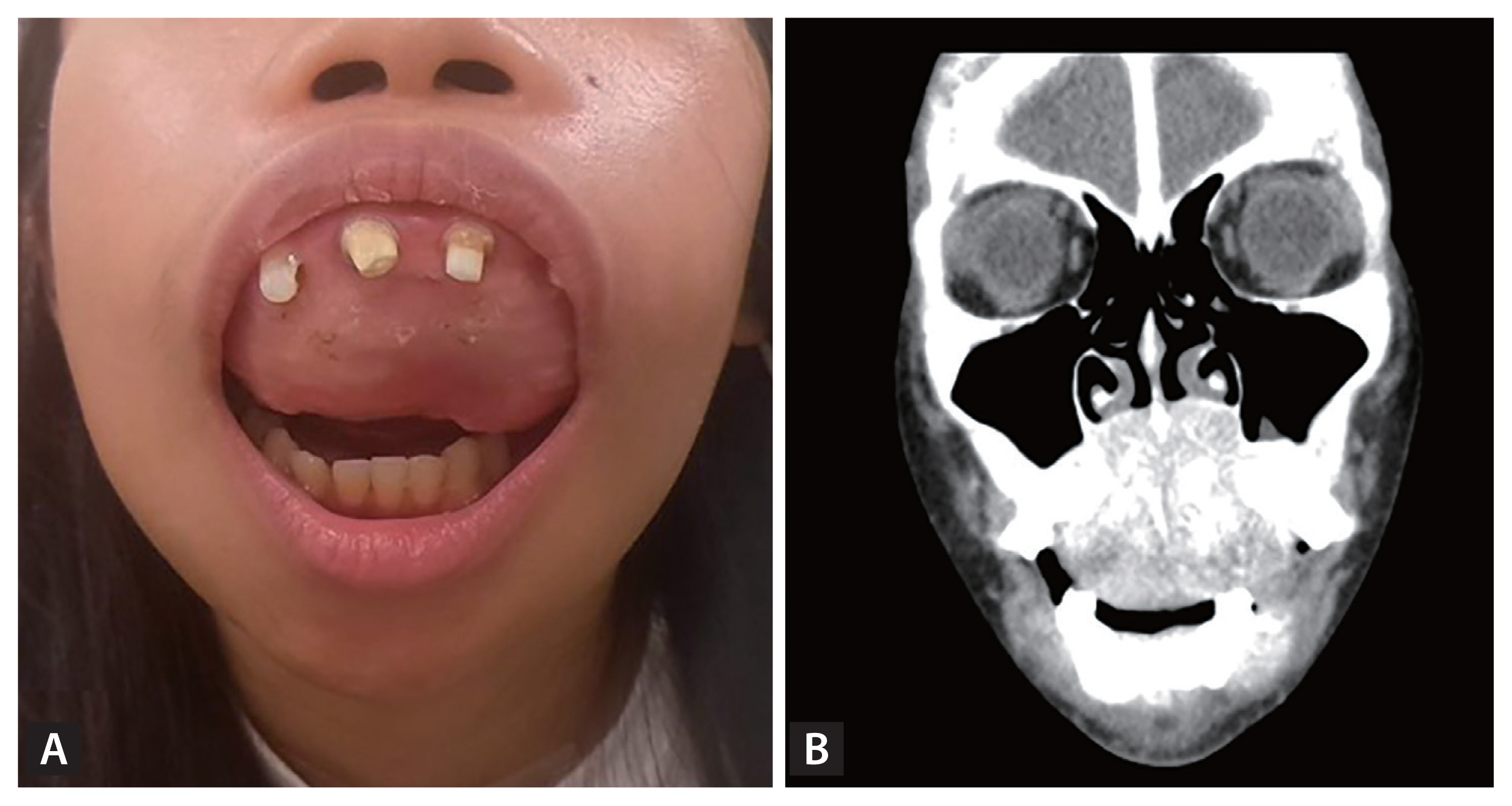
A 26-year-old woman who had been on hemodialysis for 7 years presented with a gradually swelling palatal lesion causing difficulty in phonation, dysphagia, and splaying of the maxillary teeth (Fig. 1A). Her PTH levels had been very high for 6 years. At this presentation, laboratory results showed: parathyroid hormone (PTH) level > 3,000 pg/mL, phosphorus level 6 mg/dL, alkaline phosphatase level 1,416 U/L, and normal calcium level (9 mg/dL). Computed tomography showed expansile lesions over bilateral mandibular bodies and palate with ground-glass appearance and mild osteolytic change of the left lateral sphenoid ridge (Fig. 1B). An incisional biopsy showed proliferation of fibrous tissue with a misshapen trabecular bone, scant osteoblastic rimming and mild increase in osteoclastic infiltration, compatible with fibrous dysplasia or brown tumor. Guanine nucleotide-binding protein/alpha-subunit (GNAS) exon 8 DNA sequencing showed substitution of arginine 201 with serine (R201S), confirming the diagnosis of fibrous dysplasia. She received subtotal parathyroidectomy and deceased kidney transplantation 2 months later. To correct the facial deformity, she received partial maxillectomy, resection of the maxillary tumor followed by reconstruction and tooth replantation nearly 1.5 years later. After nearly 2 years of follow-up, she had satisfactory function and aesthetic appearance (Fig. 2).
Brown tumor and fibrous dysplasia can show radiological and histopathologic similarities in patients with chronic renal failure, in whom GNAS gene analysis is a reliable and valuable adjunct to differentiate fibrous dysplasia from brown tumor. The management of patients with craniofacial fibrous dysplasia should be individualized. Fibrous dysplasia is benign, and surgical treatment is indicated in symptomatic cases with pain, fracture or significant deformity. Written informed consent was obtained from the patient.

 PDF Links
 PubReader
 ePub Link
 Full text via DOI
 Download Citation
 Print
Print


