


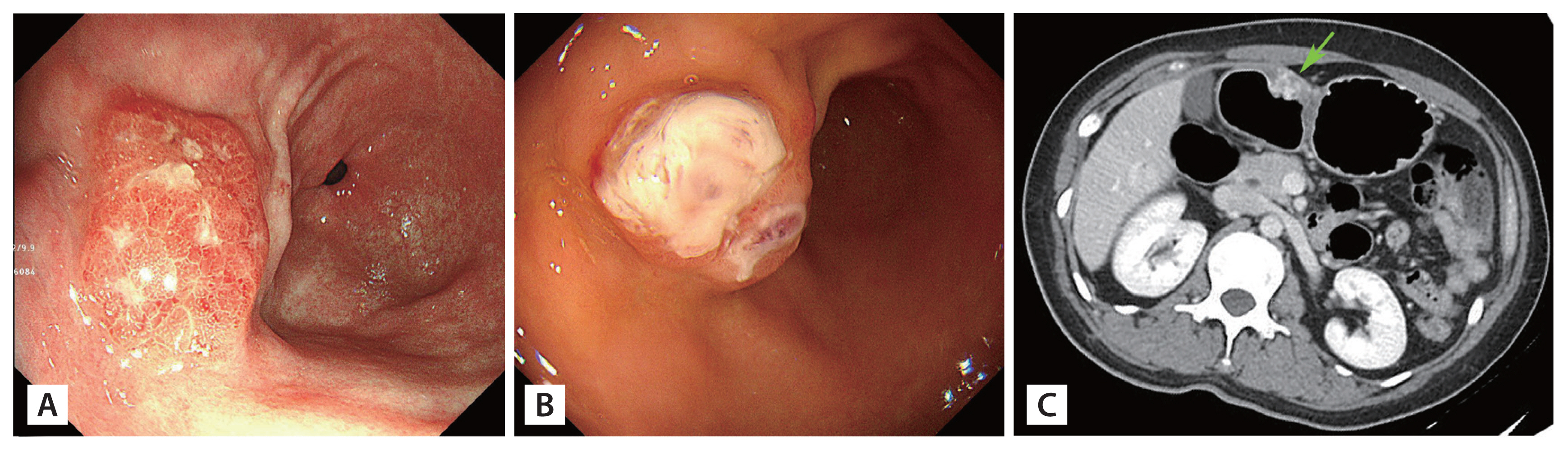
A 38-year-old woman was referred to Seoul National University Boramae Medical Center with dizziness and iron-deficiency anemia (hemoglobin 8.0 g/dL) detected during a medical check-up. Esophagogastroduodenoscopy revealed about 2-cm polypoid mass with a hyperemic surface in antrum along the anterior wall, needing to exclude early gastric carcinoma type I (Fig. 1A). Endoscopic-biopsy mucosal tissue demonstrated a tiny proliferating vascular lesion. Despite oral ferrous sulfate and intravenous iron administration weekly over 15 months, dizziness, fatigue, and anemia were aggravated (hemoglobin 5.4 g/dL). On a follow-up esophagogastroduodenoscopy, the polypoid mass progressed into a gastric ulcer (Fig. 1B). Follow-up biopsy of three times revealed just gastritis or ulceration. Melena once ensued. Proton pump inhibitors (PPIs) or non-steroidal anti-inflammatory drugs (NSAIDs)were never medicated. Stomach computed tomography showed about 1.9-cm intraluminal mass, peripheral enhancement with gradual fill-in/central pitting (Fig. 1C). Inevitably, wedge resection of the stomach was performed (Fig. 2A). Throughout the entire gastric wall, multifocal intravascular/stromal cellular micronodules mingled with vascular components, further, malformed thick-walled blood vessels (Fig. 2B); factor VIII-related antigen/CD31/CD34 (+) in vascular lining endothelium, ETS-related gene (ERG) (+) in both lining and adjacent tumor cells, human herpsevirus 8 (HHV-8) 8 (ŌłÆ), Ki-67 index, 3% immunohistochemically; and wild type isocitrate dehydrogenases (IDH1/2) by Sanger sequencing were observed. She was diagnosed with gastric composite hemangioendothelioma; composed of spindle cell/epithelioid hemangioendothelioma and vascular malformation. No recurrence occurred during the 18-month follow-up after surgical resection.
Composite hemangioendothelioma is an extremely rare vascular neoplasm of intermediate malignancy, characterized by histologically two or more distinct vascular components combination; vascular malformation, spindle/epithelioid hemangioendothelioma, and angiosarcomatoid appearance, etc. Composite hemangioendothelioma mostly affects the skin. Exceptionally rare cases in visceraŌĆöspleen and kidneyŌĆöhave been stated, but not yet reported in the stomach. Gastric composite hemangioendothelioma can cause severe iron-deficiency anemia from gastric mass bleeding, and it is crucial to secure clear surgical resection margins, and warn patients of significant potential for recurrence and close follow-up.
This study was approved by the Institutional Review Board of Seoul National University Boramae Hospital (approval #30-2020-286).

 PDF Links
 PubReader
 ePub Link
 Full text via DOI
 Download Citation
 Print
Print


