


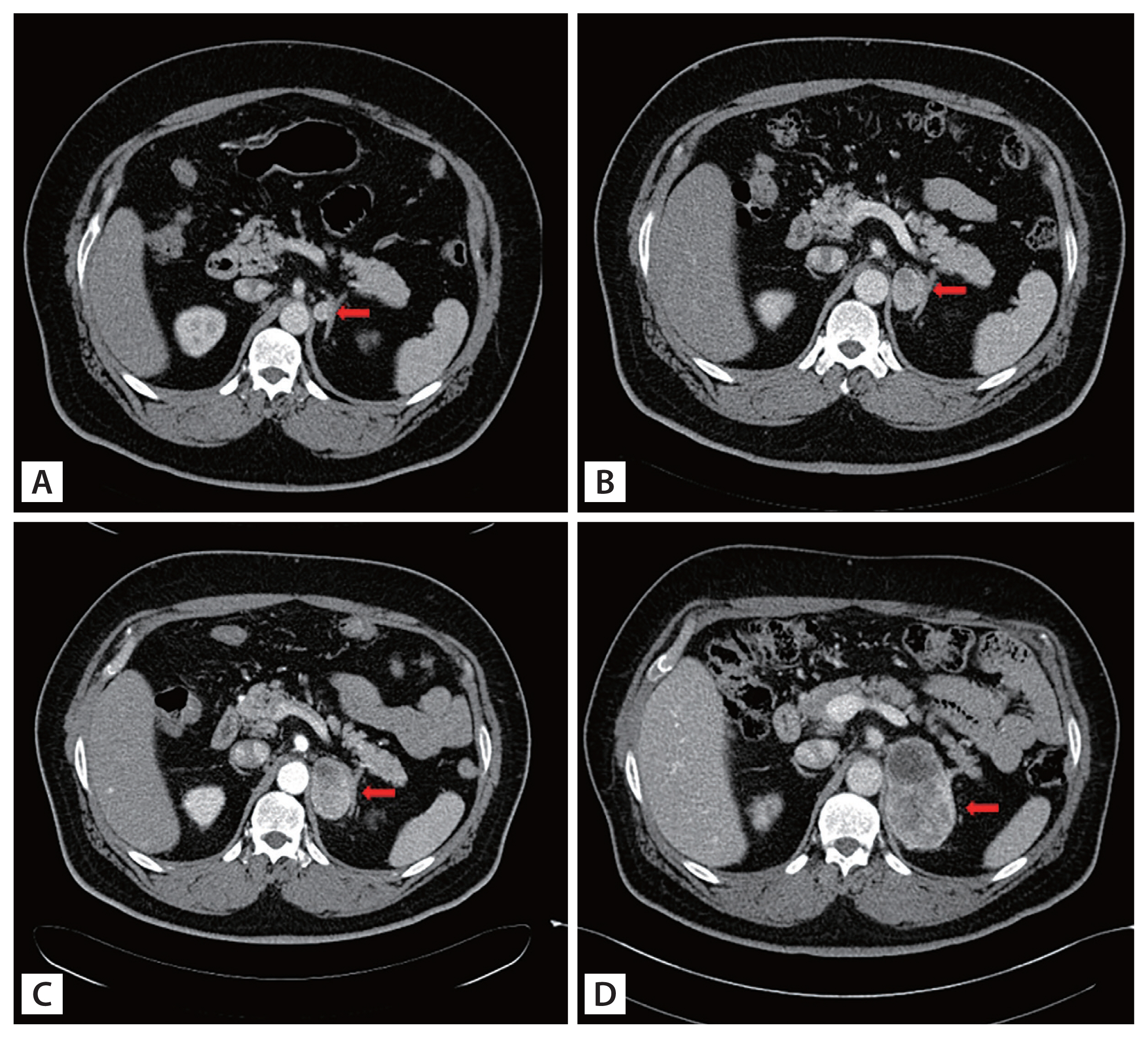
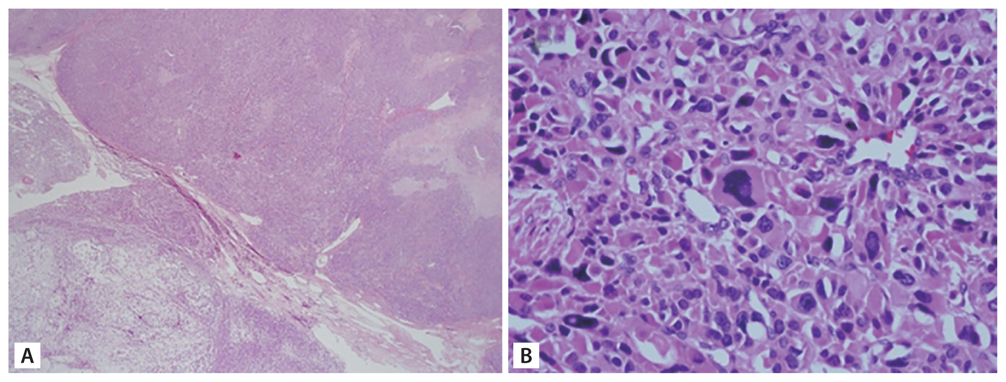
A 48-year-old man presented to our endocrinology department for the evaluation of the left adrenal incidentaloma discovered during fatty liver assessment. Abdominal computed tomography revealed a 1.8 cm left adrenal incidentaloma (Hounsfield units 40). The results of laboratory findings were as follows: serum adrenocorticotropic hormone 54.3 pg/mL, cortisol 6.39 μg/dL, renin activity 0.41 ng/mL/hr, aldosterone 12.8 ng/dL, thyroid-stimulating hormone 4.86 mIU/L, and free thyroxine 1.30 ng/dL. The 24-hour urine collection results were as follows: epinephrine 12.82 μg/day, norepinephrine 43.71 μg/day, vanillyl mandelic acid 3.10 mg/day, cortisol 39.3 μg/day. The tumor I-131 metaiodobenzylguanidine (MIBG) scan showed no abnormal uptake. The patient was diagnosed with nonfunctioning adrenal incidentaloma, and was recommended for regular follow-up. The adrenal mass continuously increased from 1.8 cm in size at baseline to 2.7 × 2.3 cm at month 15, and finally to 8.0 × 4.3 cm after 6 years of follow-up suggesting a high possibility of malignancy (Fig. 1). No hormonal change was observed during follow-up. However, the patient refused surgical management several times for personal reasons. At last, after 6 years of follow-up, he received the surgery. The pathology revealed adrenal cortical carcinoma (ACC) with 4–5/10 high-power field (HPF) mitotic rate (Fig. 2). After surgery, the patient went through several systemic chemotherapy but expired due to recurrent multiple ACC metastasis.
ACCs are rare tumors with an estimated annual incidence of 0.7 to 2 cases per million population. ACC prognosis is poor, with an overall survival of 3.21 years from diagnosis. Therefore, evaluation of adrenal incidentaloma is crucial for the early diagnosis of ACC. Even if the initial presentation of adrenal incidentaloma is small without hormonal function, intense regular follow-up is necessary to detect any malignancies. Our case highlights the natural growing course of ACC, first diagnosed as a small incidentaloma. The study was approved by Institutional Review Board of Uijeongbu St. Mary’s Hospital, the Catholic University of Korea (UC21ZISI0128).
 PDF Links
 PubReader
 ePub Link
 Full text via DOI
 Download Citation
 Print
Print


