


A 79-year-old man was referred to our hospital with hematochezia and anemia (Hb level: 8.9 g/dL). He had several comorbidities including diabetes, hypertension, dyslipidemia, and Parkinson’s disease. In addition, he had developed COVID-19 infection one month before his referral to our hospital. At the time of his admission to our hospital, generalized edema and skin petechiae were prominent, and hematochezia, diarrhea, and azotemia were gradually worsened. Abdominopelvic computed tomography showed massive ascites and diffuse bowel edema; however, a definite gastrointestinal bleeding lesion was not detected. Because of a high suspicion of COVID-19-associated systemic vasculitis, empirical intravenous methylprednisolone was administered and hemodialysis was initiated; however, massive hematochezia and anemia persisted (Hb level: 5.7 g/dL).
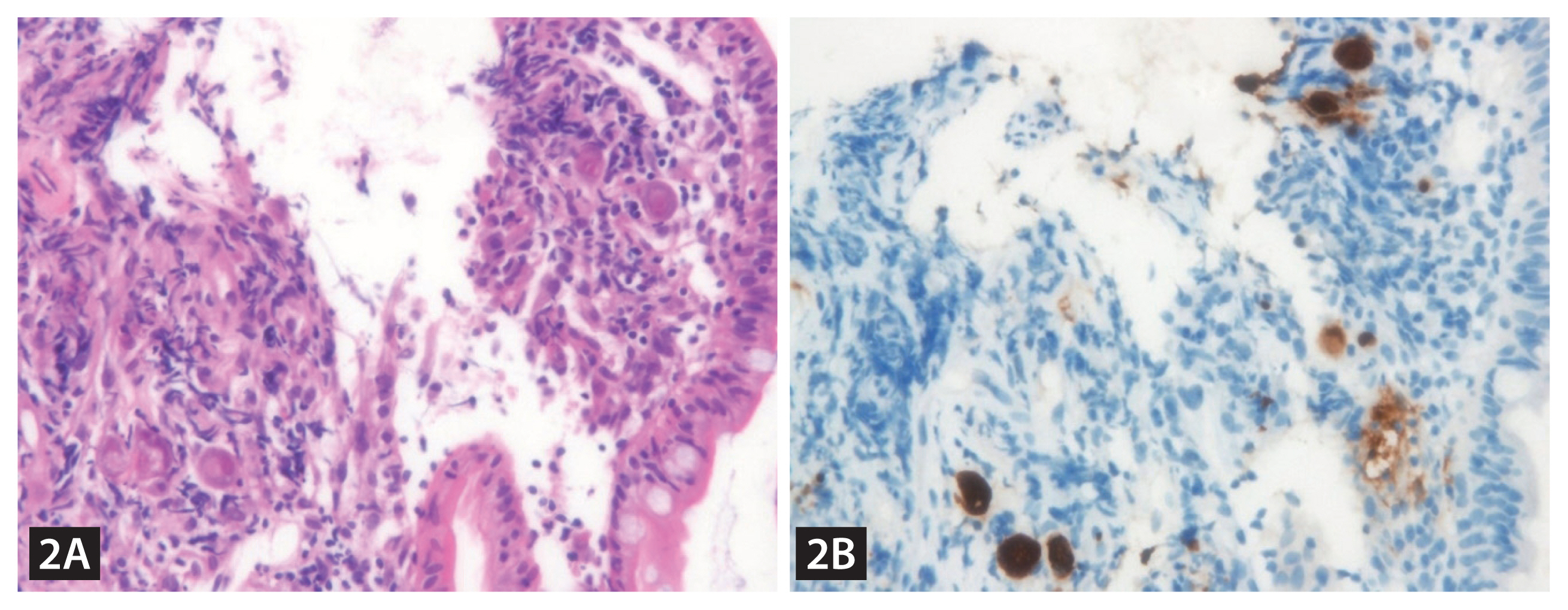
Emergent endoscopy revealed fresh blood from the terminal ileum in the lower endoscopy, whereas there was no sign of recent bleeding in the upper endoscopy. Considering a high possibility of overt small bowel bleeding and a high risk of aspiration during bowel preparation, peroral single-balloon enteroscopy was performed. It revealed multiple ulcers, mucosal friability, and hyperemia in the proximal and middle jejunum, suggesting cytomegalovirus (CMV) enteritis (Fig. 1). Thus, intravenous ganciclovir therapy was started, and a diagnosis of CMV enteritis was finally made based on histopathologic findings (Fig. 2). However, recurrent bleeding with hypotension persisted despite additional angiographic embolization for the proximal jejunum. Thus, surgical resection and anastomosis of the proximal jejunum were performed. The surgical specimen showed findings corresponding to CMV enteritis. However, pancytopenia and azotemia gradually aggravated, and the patient finally died one month after the surgery.
CMV enteritis could manifest as gastrointestinal bleeding in patients with marked ulceration. However, massive bleeding as in this case is relatively rare. The diagnosis of CMV enteritis is challenging owing to the difficulty in performing a biopsy. However, enteroscopy can facilitate early diagnosis and proper treatment.

 PDF Links
 PubReader
 ePub Link
 Full text via DOI
 Download Citation
 Print
Print


