


INTRODUCTION
Foreign body ingestion in the pediatric population usually occurs by accident. In adults, the most common causes are strictures of the gastrointestinal tract, gastrointestinal motility disorders, psychiatric disorders, mental retardation, false teeth, or impairment caused by alcohol, as well as by those seeking some secondary gain in accessing a medical facility1, 2). Most small foreign bodies will pass spontaneously through the entire alimentary tract and out in the feces. However, 10% to 20% of cases will not pass physiologic or pathologic strictures of the esophagus and will require intervention3). Removal with a flexible endoscope is preferred because of its low morbidity rate, reduced cost, and the ability to diagnose other diseases during the procedure. Foreign body impaction in the duodenum is very rare, between 0 (0%) to 5 (2.5%) in population studies4-8). Here we report the case of a clamshell impacted in the second duodenal portion, resulting in a clinical presentation mimicking a biliary stone.
CASE REPORT
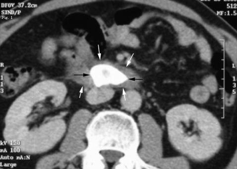
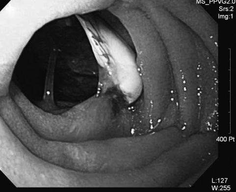
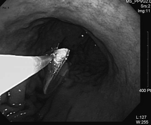
A 60-year-old man with no previous abdominal complaints, dementia, or psychological disease, presented to our outpatient internal medicine department with a one-day history of abdominal pain. The pain was initially localized to the epigastrium, weakening in intensity as time went on. There was no nausea, vomiting, febrile or chilly sensations, or diarrhea. A physical examination revealed no localized tenderness in the abdomen and a temperature of 37Ōäā. Laboratory tests indicated a slightly elevated white cell count of 11,130/mm3 with 78.5% neutrophils, elevated aspartate aminotransferase/alanine aminotransferase levels (52/44 IU/L), and normal serum amylase (86 U/L). Urinalysis and other laboratory data were within normal limits. An abdominal computed tomography scan showed a mild dilatation of the common bile duct, but no prominent obstructive lesion was seen. The computed tomography scan also showed a 3├Ś2-cm calcific shadow in the second portion of the duodenum (Figure 1). An abdominal ultrasonography revealed an echogenic lesion in the duodenum (Figure 2). An arch-like echogenic rim and posterior acoustic shadowing of the lesion gave it the appearance of a stone originating from the biliary tree. Upper gastrointestinal endoscopy revealed a broken piece of clamshell in the second portion of the duodenum (Figure 3). Endoscopic retrieval of the clamshell by a Dormia Basket was performed safely (Figure 4, 5) and the patient was discharged neventfully on the day of the procedure.
DISCUSSION
Ingestion of a foreign body is commonly encountered in children, adults with intellectual impairment, psychiatric illness or alcoholism, and elderly patients with dental prosthetics9, 10), but foreign body impaction in the duodenum is rare. According to a large study in China by Li et al11), a duodenal foreign body was reported in only 50 cases (4.6%) among the 1088 cases with foreign bodies in the upper GI tract. Most of these cases involved small, smooth objects, such as metallic pieces and glass balls. In general, objects wider than 2 cm do not pass through the pylorus and tend to lodge in the stomach, while objects longer than 5 cm tend to get caught in the duodenal sweep12, 13). The broken piece of clamshell in the present case was 2 cm in diameter and 3 cm in length, with a pointed edge. Objects that lodge in the gastric lumen often remain there for long periods without adverse consequences14). Watchful waiting is generally justified and may include administration of emetics, laxatives, or spasmolytics, depending on the type and site of object12). However, perforation is always a potential complication when sharp objects are ingested. Sharp objects that lodge in the same place for more than 2 to 3 days15) or objects in the stomach that have not moved for more than 5 to 6 days16) are unlikely to pass and should be removed endoscopically. Up to 15% to 35% of sharp and pointed foreign bodies ingested penetrate the wall of the gastrointestinal tract17) and should be removed by gastrostomy18). A sharp or pointed foreign body in the stomach or duodenum found during an endoscopic evaluation should be removed, even if the patient is asymptomatic. Usually, adults that ingest pointed objects are prisoners or psychiatric patients, and carry a higher complication rate and surgical rate than for accidental ingestion13).
In this case, the patient had no psychological disease and was not aware that he had eaten the clamshell. As a result, the diagnosis before performing upper gastrointestinal endoscopy was a gall stone in the duodenum, based on abdominal imaging. Endoscopic removal of foreign bodies requires skilled endoscopists and accessories such as snares, dormia baskets, or strong-toothed graspers. Commercial accessories developed specifically for removing foreign objects are also available-for example, soft-latex protector hoods19) and overtubes. Endoscopic retrieval of sharp objects is accomplished with the retrieval forceps (rat-tooth, biopsy, or alligator jaws) or a snare. The risk of mucosal injury during sharp-object retrieval can be minimized by orienting the object with the point trailing during extraction with an overtube. In this case, the small internal diameter of the overtube (11 to 15 mm) prevented the removal of the clamshell. Improved diagnostic and therapeutic modalities can now reduce the rate of morbidity and mortality associated with foreign body ingestions. However, it appears that the clinician must still maintain a high degree of suspicion and a prudent management plan when the possibility of foreign body impaction in the duodenum exists.


 PDF Links
 PubReader
 ePub Link
 Full text via DOI
 Download Citation
 Print
Print


