Korean J Intern Med > Volume 16(2); 2001 > Article
Kim, Oh, Kim, Choi, Kim, Baik, Choi, and Lee: Fra-1 Expression in Malignant and Benign Thyroid Tumor

Abstract

Background:

The differential diagnosis of thyroid nodules is very important in deciding the treatment modality and the fine needle aspiration is the best diagnostic method. But, there are some limitations in use because of inadequate test materials and difficulty in interpreting. According to the study of oncogene and tumor suppressor gene about the origin of thyroid tumor, expression of Fra-1, one of AP-1 complex, is increased in thyroid neoplasm, though not present in the normal tissue. So, there is a possibility that it will be used as a method for the differential diagnosis of thyroid nodules. We tried to know whether presence or absence of Fra-1 expression can be used as a diagnostic method in differential diagnosis of thyroid nodules using the immunohistochemical (IHC) staining method.

Method:

In 4 types of thyroid tumor that were confirmed by histologic diagnosis after operation (18 cases of adenomatous goiter, 16 cases of follicular adenoma, 30 cases of papillary cancer, 10 cases of follicular cancer), IHC staining method was performed to evaluate the expression of Fra-1.

Result:

In papillary and follicular thyroid cancers, the expression of Fra-1 was stronger than in benign thyroid tumor, but there was no difference in Fra-1 expression between the two types of carcinoma. Weak expression of Fra-1 was observed in all cases of follicular adenoma, though it was weaker than in carcinoma, and it was also weakly expressed only in some cases (33%) of adenomatous goiter.

Conclusion:

The expression of Fra-1 was stronger in thyroid cancer than in benign thyroid tumor, but it was impossible to differentiate thyroid cancer from benign thyroid tumor by presence or absence of Fra-1 expression using IHC staining method.

INTRODUCTION

As a very common endocrine disease, thyroid nodules are occurred by 1–7% according to the iodine uptake1,2) and their interpretation, whether malignant or benign, is a very important factor in treatment. The methods of differential diagnosis are clinical manifestation of thyroid nodules, ultrasonographic finding, isotope scanning finding and fine needle aspiration, of which the fine needle aspiration test is best for a cancer diagnosis with 85% of sensitivity and 90–95% of specificity3). But, there are limitations on use because an inadequate or improper specimen often obstructs diagnosis and, moreover, it has a very poor sensitivity for a diagnosis of follicular thyroid cancer. As for the origin of thyroid cancer, various oncogenes and tumor suppressor genes are being studied, but it is still difficult to differentiate thyroid cancer from benign thyroid tumor by clinical application of them46). Fra-1 is one of 4 Fos family (c-Fos, Fos B, Fra-1, Fra-2) forming AP-1 complex and it is known that its activity increases at a level of mRNA when transformed into a tumor cell, as well as in the test using thyroid cancer culture cell line7,8). According to the study using the pathologic thyroid tissue, expression of Fra-1 is absent in normal thyroid tissue, whereas it is present in thyroid cancer tissue, which suggested a possibility of use as a differential diagnosis of benign tumor and malignant tumor8). For a differential diagnosis of thyroid nodules, this study compared the expression of Fra-1 by the immunohistochemical (IHC) staining method in malignant thyroid tumor types, including papillary and follicular thyroid cancer, and benign thyroid tumor types, including adenomatous goiter and follicular adenoma.

MATERIALS AND METHODS

1. Subjects

4 types of thyroid tumor (18 cases of adenomatous goiter, 16 cases of follicular adenoma, 30 cases of papillary cancer and 10 cases of follicular cancer) confirmed by histologic diagnosis after operation were researched.

2. Methods

The expression of Fra-1 was measured by the immunohistochemical (IHC) staining method. First, the prepared paraffin tissue was put on a slide in a thickness of 4 μm and dried for over 2 hours at room temperature. It was washed in Xylene for 5 min. 3 times and then in alcohol of 100%, 95%, 80% for 2 min, in turn, to remove the paraffin. The slide was immersed in a 0.1 M Sodium citrate buffer and put in a microwave oven for 5 min. 2 times to expose the antigenecity, and then stabilized at room temperature. It was purified in a solution of pure methanol and 3% hydrogen peroxide mixed by 4:1 for 30 min to remove endogenous hydrogen peroxide. After the blocking antibody was treated for 30 min., it underwent the rabbit polyclonal Fra-1 (Santa Cruz, USA) as the primary antibody before incubation at 4°c for one night and then biotinylated goat anti-rabbit IgG (Vectastatin ABC kit, Vector, USA) as the secondary antibody for 30 min at room temperature. Thereafter it was reacted with ABC reagent for 30 min, stained with DAB (diaminobenzidin, DAKO, USA) for 5 min. and passed through hematoxylene control staining. After the dehydration course of alcohol and Xylene washing, it was made as a permanent slide with Permount solution. The expression of Fra-1 was evaluated by 4 scales from 0 to 3, which means no staining for 0 and the highest degree of staining for 3.

3. Statistics

For statistical result, SPSS for window 7.5 was used and ANOVA was executed to compare the expression of Fra-1, which judged p <0.05 as significant.

RESULTS

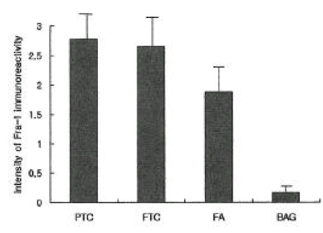
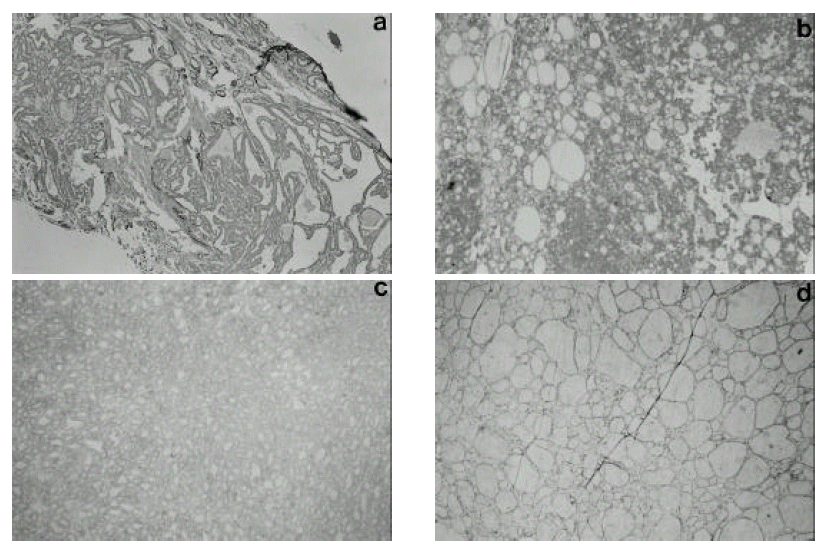
The expression of Fra-1 was stronger in papillary and follicular cancer than in benign thyroid tumor, but there was no difference in Fra-1 expression between the two types of carcinoma. Weak expression of Fra-1 was observed in all cases of follicular adenoma, and it was also weakly expressed in 6 out of 18 (33%) cases of adenomatous goiter (Figure 1).

DISCUSSION

AP-1 complex consists of 3 types of Jun family (c-Jun, Jun B, Jun D) and 4 types of Fos family (c-Fos, Fos B, Fra-1, Fra-2) and is related with activation of lots of genes, concerning regulation of cell-proliferation, and tumorigenesis, transformation by forming various kinds of homoor hetero-dimers9,10). As to tumorigenesis, the function of AP-1 complex became known for the first time when retro virus having V-jun and V-fos oncogenes was isolated and the function of oncogenes like c-Jun, Jun B, c-Fos, Fos B was clarified1114). On the other hand, it was reported that Jun-D negatively regulates fibroblast growth and partially antagonizes transformation by ras15). Some researches have been made about the function of AP-1 in transformation of mouse 3T3 and rat embryo fibroblast1618), and another research revealed that cultured cell from c-Jun-defective mouse resists against transformation by ras19). With regard to the role of AP-1 in the transformation of epithelial cell, the thyroid cell was used for the study7,8,2528) because increased AP-1 activity is essential for transformation of mouse epidermal cell2024), and thyroid cell transformation is a good model for neoplasm of epithelial cells. Among them, the test using rat thyroid culture cell line, including FRTL-5 and PC C1 3, reported that increment of AP-1 activity reflects the various changes of cell formation and Jun B and Fra-1 activity of AP-1 complex increases at a level of mRNA7). This increased Jun B and Fra-1 activity was known as being related to HMGI protein, which is known to present higher expression with malignant transformation of thyroid culture cells7,29). That is, among 3 HMGI proteins (HMGI, HMGY, HMGI-C), expression of HMGI-C protein is essential for Jun B and Fra-1 gene induction, which was suppressed in the cell line expressed anti-sense HMGI-C. When Fra-1 was suppressed by Fra-1 antisense RNA vector, malignant expression of transformed thyroid cells was reduced. So, Fra-1 gene was reported as important in cellular transformation pathways7). Seeing that regulation of Fra-1 transcription is highly dependant on AP-1 reactive inducer in the first intron of a gene and Fra-1 activity decreases in c-Fos or c-Jun deficient fibroblast, but increases by over-expression of another AP-1 family like FosB or c-Jun30), it can imply that increased Fra-1 expression involving oncogene is related to the activation of another AP-1 component made in the early stage of formation of transformation expression. Battista, et al.8) suggested that Fra-1 test using IHC staining method can be a help to differentiate thyroid cancer from benign thyroid tumor because Fra-1 stain was abundant in thyroid cancer but abscent in normal thyroid tissue from a result of study using IHC staining method to check the difference of Fra-1 expression, which was confirmed again by molecular biology technique and AP-1, especially Fra-1, c-Jun, Jun D, activity was also increased in cultured human thyroid cancer cells. This study was performed to evaluate if it is useful for a differential diagnosis of thyroid nodules by comparing the expression of Fra-1 between malignant thyroid tumor types, including papillary and follicular thyroid cancers, and benign thyroid tumor types, including adenomatous goiter and follicular adenoma. The expression of Fra-1 was much stronger in malignant thyroid tumor than in benign thyroid tumor, but there was no difference in Fra-1 expression between two types of carcinoma. In all the cases of follicular adenoma, the expression of Fra-1 was observed, though weak compared to thyroid cancers. In 6 cases out of 18 (33%) adenomatous goiter, Fra-1 was very weakly expressed. So the result revealed no specific staining reaction to the malignant thyroid tumor only, and it was not adequate to differentiate by the expression of Fra-1 whether malignant or benign the thyroid nodules are, as shown in the research of Battista, et al8). This is because the expression of Fra-1 can be essential for morphologic deformity, but not satisfactory for causing irreversible degeneration into a tumor, as mentioned in the experiment using rat fibroblast and thyroid culture cell by Bergers31) and Vallone, et al.32). In Fra-1 activity during the progression of neoplastic transformation, it is known that Fra-1 is a transcriptional goal of c-Fos and a temporary activity of c-Fos is essential for irreversible activity of Fra-17). The experimental model of skin tumor formation displayed the development of papilloma at c-Fos deficient mouse, but no progression of skin tumors33). Therefore, the possibility of a thyroid tumor induction at c-Fos deficient rat model and the research about the expression of Fra-1 at this time could be very helpful to clarify the role of Fra-1 in thyroid cancers. As a result, the expression of Fra-1 was stronger in thyroid cancer than in benign tumor, but IHC staining method appeared as irrelevant to differentiate malignant from benign tumors by the expression of Fra-1.

Figure 1.
Intensity of positive immunostaining of Fra-1 in papillary thyroid cancer (PTC), follicular thyroid cancer (FTC), follicular adenoma (FA), benign adenomatous goiter (BAG), as evaluated on a scale of 0–3 (0 and 3 corresponded to the absence and the highest degree of staining, respectively).
kjim-16-2-93-8f1.gif
Figure 2.
Immunohistochemical staining of Fra-1 in thyroid tumor, a) Strong immunoreactivity was observed in papillary thyroid carcinoma. b) Strong immunoreactivity was observed in follicular thyroid carcinoma. c) Weak immunoreactivity was observed in follicular adenoma. d) No immunoreactivity was observed in adenomatous goiter.
kjim-16-2-93-8f2.gif

REFERENCES

1. Werk EE Jr, Vernon BM, Gonzalez JJ, Ungaro PC, McCoy RC. Cancer in thyroid nodules. A community hospital survey. Arch Intern Med 144:474–4761984.
crossref pmid
2. Vander JB, Gaston EA, Dawber TR. The significance of nontoxic thyroid nodules. Final report of a 15-year study of the incidence of thyroid malignancy. Ann Intern Med 69:537–5401968.
crossref pmid
3. Caruso D, Mazzaferri EL. Fine needle aspiration biopsy in the management of thyroid nodules. Endocrinologist 1:194–2021991.
crossref
4. Farid NR, Shi Y, Zou M. Molecular basis of thyroid cancer. Endocr Rev 15:202–2321994.
crossref pmid
5. Fagin JA. Genetic basis of endocrine disease 3: Molecular defects in thyroid gland neoplasia. J Clin Endocrinol Metab 75:1398–14001992.
crossref pmid
6. Herrmann MA, Hay ID, Bartelt DH Jr, Ritland SR, Dahl RJ, Grant CS, Jenkins RB. Cytogenetic and molecular genetic studies of follicular and papillary thyroid cancers. J Clin Invest 88:1596–16041991.
crossref pmid pmc
7. Vallone D, Battista S, Pierantoni GM, Fedele M, Casalino L, Santoro M, Viglietto G, Fusco A, Verde P. Neoplastic transformation of rat thyroid cells requires the junB and fra-1 gene induction which is dependent on the HMGI-C gene product. EMBO J 16:5310–53211997.
crossref pmid pmc
8. Battista S, Nigris F, Fedele M, Chiappetta G, Scala S, Vallone D, Pierantoni GM, Mega T, Santoro M, Viglietto G, Verde P, Fusco A. Increase in AP-1 activity is a general event in thyroid cell transformation in vitro and in vivo. Oncogene 17:377–3851998.
crossref pmid
9. Angel P, Karin M. The role of Jun, Fos and the AP-1 complex in cell-proliferation and transformation. Biochim Biophys Acta 1072:129–1571991.
crossref pmid
10. Karin M, Liu Z, Zandi E. AP-1 function and regulation. Curr Opin Cell Biol 9:240–2461997.
crossref pmid
11. Schutte J, Minna JD, Birrer MJ. Deregulated expression of human c-jun transforms primary rat embryo cells in cooperation with an activated c-Ha-ras gene and transforms rat-1a cells as a single gene. Proc Natl Acad Sci USA 86:2257–22611989.
crossref pmid pmc
12. Van Amsterdam Jr, Wang Y, Sullivan RC, Zarbl H. Elevated expression of the junB proto-oncogene is essential for v-fos induced transformation of Rat-1 cells. Oncogene 9:2969–29761994.
pmid
13. Miller AD, Currant T, Verma IM. c-fos protein can induce cellular transformation: a novel mechanism of activation of a cellular oncogene. Cell 36:51–601984.
crossref pmid
14. Wisdom R, Yen J, Rashid D, Verma IM. Transformation by FosB requires a trans-activation domain missing in FosB2 that can be substituted by heterologous activation domains. Genes Dev 6:667–6751992.
crossref pmid
15. Pfarr CM, Mechta F, Spyrou G, Lallemand D, Carillo S, Yaniv M. Mouse JunD negatively regulates fibroblast growth and antagonizes transformation by ras. Cell 76:747–7601994.
crossref pmid
16. Suzuki T, Murakami M, Onai N, Fukuda E, Hashimoto Y, Sonobe MH, Kameda T, Ichinose M, Miki K, Iba H. Analysis of AP-1 function in cellular transformation pathways. J Virol 68:3527–35351994.
crossref pmid pmc
17. Mechta F, Lallemand D, Pfarr CM, Yaniv M. Transformation by ras modifies AP1 composition and activity. Oncogene 14:837–8471997.
crossref pmid
18. Vandel L, Montreau N, Vial E, Pfarr CM, Binetruy B, Castellazzi M. Stepwise transformation of rat embryo fibroblasts: c-Jun, JunB, or JunD can cooperate with Ras for focus formation, but a c-Jun-containing heterodimer is required for immortalization. Mol Cell Biol 16:1881–18881996.
crossref pmid pmc
19. Johnson R, Spiegelman B, Hanahan D, Wisdom R. Cellular transformation and malignancy induced by ras require c-jun. Mol Cell Biol 16:4504–111996.
crossref pmid pmc
20. Domann FE, Levy JP, Birrer MJ, Bowden GT. Stable expression of a c-JUN deletion mutant in two malignant mouse epidermal cell lines blocks tumor formation in nude mice. Cell Growth Differ 5:9–161994.
pmid
21. Dong Z, Birrer MJ, Watts RG, Matrisian LM, Colburn NH. Blocking of tumor promoter-induced AP-1 activity inhibits induced transformation in JB6 mouse epidermal cells. Proc Natl Acad Sci USA 91:609–6131994.
crossref pmid pmc
22. Rosenberger SF, Bowden GT. Okadaic acid stimulated TPE binding activity in a papilloma producing mouse keratinocyte cell line involves increased AP-1 expression. Oncogene 12:2301–23081996.
pmid
23. Li JJ, Dong Z, Dawson MI, Colburn NH. Inhibition of tumor promoter-induced transformation by retinoids that trans-repress AP-1 without transactivating retinoic acid response element. Cancer Res 56:483–4891996.
pmid
24. Joseloff E, Bowden GT. Regulation of the transcription factor AP-1 in benign and malignant mouse keratinocyte cells. Mol Carcinog 18:26–361997.
crossref pmid
25. Fusco A, Portella G, Fiore PP, Berlingieri MT, Di Lauro R, Schneider AB, Vecchio G. A mos oncogene-containing retrovirus, myeloproliferative sarcoma virus, transforms rat thyroid epithelial cells and irreversibly blocks their differentiation pattern. J Virol 56:284–2921985.
crossref pmid pmc
26. Fusco A, berlingieri MT, Di Fiore PP, Portella G, Grieco M, Vecchio G. One- and two-step transformations of rat thyroid epithelial cells by retroviral oncogenes. Mol Cell Biol 7:3365–33701987.
crossref pmid pmc
27. Berlingieri MT, Pertella G, Grieco M, Santoro M, Fusco A. Cooperation between the polyomavirus middle-T-antigen gene and the human c-myc oncogene in a rat thyroid epithelial differentiated cell line: model of in vitro progression. Mol Cell Biol 8:2261–22661988.
crossref pmid pmc
28. Berlingieri MT, Santoro M, Battaglia C, Grieco M, Fusco A. The adenovirus E 1A gene blocks the differentiation of a thyroid epithelial cell line: however the neoplastic phenotype is achieved only after cooperation with other oncogenes. Oncogene 8:249–2551993.
pmid
29. Berlingieri MT, Manfioletti G, Santoro M, Bandiera A, Visconti R, Giancotti V, Fusco A. Inhibition of HMGI-C protein synthesis suppresses retrovirally induced neoplastic transformation of rat thyroid cells. Mol Cell Biol 15:1545–15531995.
crossref pmid pmc
30. Bergers G, Graninger P, Braselmann S, Wrighton C, Busslinger M. Transcriptional activation of the fra-1 gene by AP-1 is mediated by regulatory sequences in the first intron. Mol Cell Biol 15:3748–37581995.
crossref pmid pmc
31. Bergers G, Graninger P, Braselmann S, Wrighton C, Busslinger M. Transcriptional activation of the fra-1 gene by AP-1 is mediated by regulatory sequences in the first intron. Mol Cell Biol 15:3748–37581995.
crossref pmid pmc
32. Vallone D, Battista S, Pierantoni GM, Fedele M, Casalino L, Santoro M, Viglietto G, Fusco A, Verde P. Neoplastic transformation of rat thyroid cells requires the junB and fra-1 gene induction which is dependent on the HMGI-C gene product. EMBO J 16:5310–53211997.
crossref pmid pmc
33. Saez E, Rutberg SE, Mueller E, Oppenheim H, Smoluk J, Yuspa SH, Spiegelman BM. c-fos is required for malignant progression of skin tumors. Cell 82:721–7321995.
crossref pmid
TOOLS
METRICS Graph View
  • 12 Crossref
  • 14 Scopus
  • 9,328 View
  • 57 Download
Related articles

Editorial Office
101-2501, Lotte Castle President, 109 Mapo-daero, Mapo-gu, Seoul 04146, Korea
Tel: +82-2-2271-6792    Fax: +82-2-790-0993    E-mail: kaim@kams.or.kr                

Copyright © 2024 by Korean Association of Internal Medicine.

Close layer
prev next