Disseminated Cryptococcosis in a Patient with Pituitary Cushing’s Disease
Article information
Abstract
Disseminated cryptococcosis mainly occurs in patients with cell-mediated immunity disorders. A case of disseminated cryptococcosis, in a patient with pituitary Cushing’s disease, is reported. Cultures of blood, cerebrospinal fluid (CSF) and aspirates of a skin lesion all grew Cryptococcus neoformans. Despite antifungal treatment, with amphotericin-B, the patient died within 3 weeks.
INTRODUCTION
Cushing’s syndrome is characterized by supraphysiological cortisol levels, and may be exogenous, due to steroid therapy, or endogenous when secondary to a functioning tumor of the pituitary or adrenal glands1). Regardless of etiology, all cases of endogenous Cushing’s syndrome are due to an increased production of cortisol by the adrenal gland. Excessive corticosteroid impairs the cell-mediated immunity, and therefore, patients with Cushing’s syndrome are prone to infection2–4). There have been several reports of cryptococcosis in patients with endogenous Cushing’s syndrome2, 4, 5), but most of these have hyperproduction of corticosteroid due to ectopic ACTH syndrome or adrenal tumors. Moreover, most cases are the pulmonary or meningeal forms of cryptococcosis, with disseminated cryptococcosis rarely reported. In this report, a case of rapidly fatal, disseminated cryptococcosis, in a patient with pituitary Cushing’s disease, is reported.
CASE REPORT
A 49-year-old woman was admitted to Seoul National University Hospital, due to a headache and fever. Her past medical history included type 2 diabetes mellitus, essential hypertension and osteoporosis. A compression fracture of the lumbar spine had been diagnosed 1 year prior to her admission, for which she had been in a nearly bed-ridden state. In the 2 weeks prior to her admission, she developed a sudden headache and fever, which was treated with antipyretics and analgesics. However, her symptoms did not improve, and her mental status deteriorated. The patient was transferred to our hospital.
A physical examination showed a temperature of 38.2°C, a blood pressure of 170/100 mmHg, a pulse rate of 102 beats/min, and normal respirations. The patient had a moon face, truncal obesity and neck stiffness. On oscultation of the lungs, there were fine crackles in the left lower lung field. An abdominal examination revealed no remarkable findings. The patient had a painful erythematous bullous lesion in the left leg, but doppler ultrasonography of the lower extremity showed no evidence of deep vein thrombosis.
The values obtained during the initial laboratory examination were as follows: WBC count 9,450 cells/mm3, hemoglobin 11.4 g/dL, hematocrit 32.6%, platelet count 230,000 cells/mm3, sodium 126 mmol/l, potassium 2.6 mmol/l, and creatinine 0.7 mg/dL. The liver function tests, and prothrombin and partial thromboplastin times, were all within normal limits. A chest radiograph showed infiltrates and consolidation of the left lung. An examination of the cerebrospinal fluid (CSF) showed no red (RBC) or white blood cells (WBC), a protein level of 118 mg/dL and a glucose level of 50 mg/dL. A gram stain for CSF showed many encapsulated yeasts. Cultures of the aspirate of the erythematous bullous lesion in the leg, and blood and CSF samples, obtained at the time of admission, yielded Cryptococcus neoformans.
She was treated with intravenous amphotericin-B at a dose of 0.6 mg/kg/day, and was investigated for the presence of an underlying immunocompromised state, especially for Cushing’s syndrome. She had no evidence of liver cirrhosis, and the serology for the human immunodeficiency virus was negative. The level of cortisol in her 24-hour urine was raised, with a value of 385.7 μg/24 hours (normal 39–200). Her serum ACTH concentration was also raised, with a level of 230 pg/ml (normal 0–60). A brain MRI revealed an extensive pituitary mass lesion, involving the sellar fossa, cavernous sinus and skull base, which extended to the suprasellar area and sphenoid sinus (Figure 1). In addition to intravenous amphotericin-B, cortisol lowering therapy, with ketoconazole and octreotide, was initiated. To further evaluate the consolidation of the left lung, a chest CT was performed. This showed segmental, or mass-like, consolidations in the posterior segment of the left upper lobe left lower lobe, and the right middle lobes, which were combined with small nodules, with ground-glass opacity. Despite antifungal treatment, with amphotericin-B, repeated cultures of the CSF and the wound, which were performed on the 14th -hospital day, still grew Cryptococcus neoformans. Despite 15 days of the antifungal therapy the patient died on the 19th day of hospitalization.
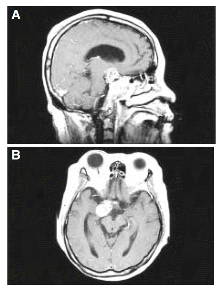
Sagittal view (A) and transverse view (B) of the brain MRI show an extensive pituitary mass lesion, involving the sella fossa, cavernous sinus and skull base, which extended to the suprasellar area and sphenoid sinus.
DISCUSSION
Corticosteroid therapy impairs the immunity, and predisposes the patient to various viral, bacterial, fungal and parasitic infections3). Patients with endogenous Cushing’s syndrome can have the same spectrum of infections as occurring in patients treated with corticosteroids2). The extent of immunological impairment and the risk of infection correlate with the degree of cortisol excess2). An opportunistic infection is therefore less likely to occur in patients with pituitary Cushing’s disease than in those with greater levels of corticosteroid overproduction due to adrenal tumors or ectopic ACTH syndrome2). In this report, it is noted that disseminated cryptococcosis developed in a patient with pituitary Cushing’s disease.
Cryptococcus neoformans is an encapsulated yeast, found ubiquitously in soil. Despite the high prevalence of the organism in the environment, human cryptococcal infections are rare, with the exception of patients with cell-mediated immunity disorders, such as AIDS, and organ transplantation, or in patients undergoing corticosteroid therapy6). The organs most commonly affected by Cryptococcus species are the CNS and lungs. Disseminated cryptococcosis has been reported in patients with severe impairment of their cell-mediated immunity, such as liver cirrhosis7), acute leukemia8), solid organ transplantion9), or AIDS10). To our knowledge, there are no reports of disseminated cryptococcosis in patients with endogenous Cushing’s syndrome.
This case emphasizes that cryptococcosis can be rapidly fatal, with multi-organ dissemination, in patients with endogenous Cushing’s syndrome, even in pituitary Cushing’s disease. Extreme hypercortisolism is an immunological endocrine emergency. Pharmacological and surgical treatment for lowering the plasma cortisol concentrations, to physiological ranges, should be promptly initiated.