Radiation nephritis: 99mTc hydroxydiphosphonate bone scan, 99mTc dimercaptosuccinic acid renal scan, and 18F-FDG PET/CT findings
Article information
A 20-year-old male had Ewing’s sarcoma of the right paraspinal region (T12–L2 vertebra level) and was treated with chemotherapy and radiation therapy (RT). 99mTc hydroxydiphosphonate bone scan was performed 4 months after the end of treatment. Intense uptake was noted in both kidneys (Fig. 1). On the fluorodeoxyglucose (FDG) positron emission tomography/computed tomography (PET/CT) for response evaluation, mild uptake was also seen in both kidneys (Fig. 2A and 2B). On 99mTc dimercaptosuccinic acid (DMSA) scan, photon defects were noted in both kidneys (Fig. 2C). When we reviewed the RT plan, the medial aspect of both kidneys was included in the radiation field (Fig. 2D and 2E). On contrast-enhanced CT, low-densities were noted in both kidneys (Fig. 2F). We thus diagnosed radiation nephritis (RN). On 6-month follow-up bone scan, the previously noted intensity on both kidneys was decreased.
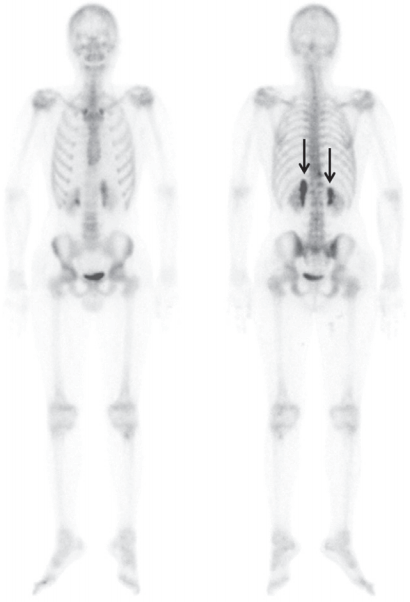
On 99mTc hydroxydiphosphonate bone scan 4 months after radiotherapy for evaluation of metastasis, intense linear band-like uptake was noted in the medial aspect of both kidneys (arrows).
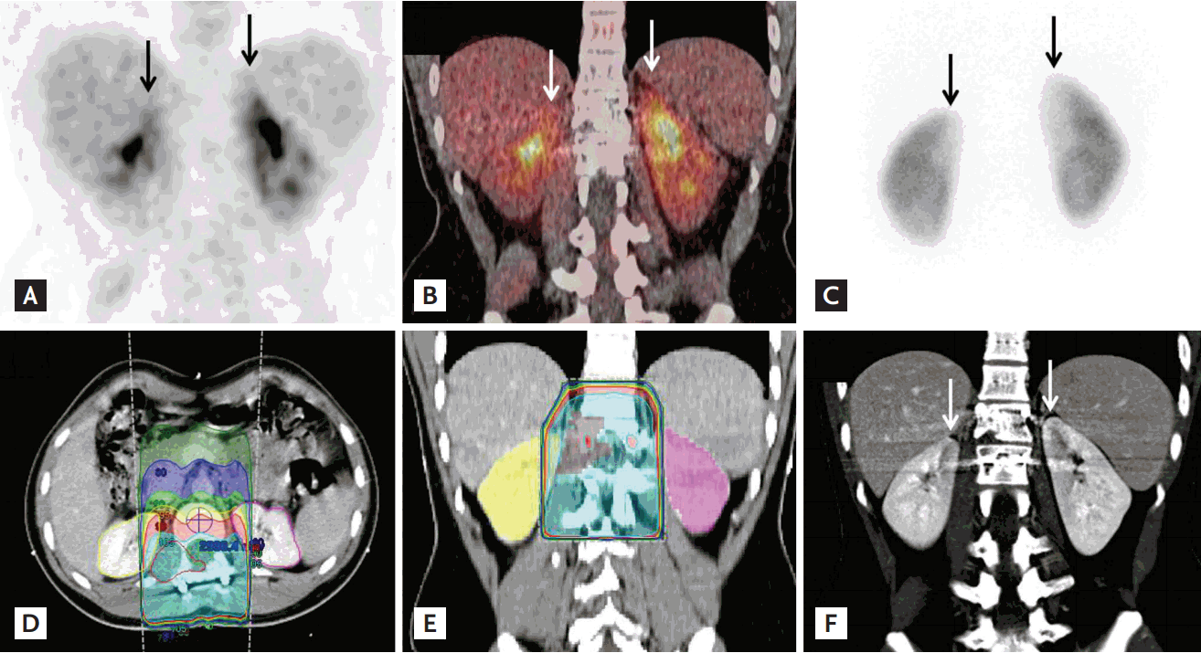
On the same day with bone scan, fluorodeoxyglucose (FDG) positron emission tomography/computed tomography (PET/CT) was performed. (A, B) Mild longitudinal uptake was seen in the medial aspect of both kidneys on 18F-FDG PET/CT (arrows). (C) Five days later, 99mTc dimercaptosuccinic acid scan was performed to evaluate functional impairment, and photon defects were noted in the medial aspect of the renal cortices. Urea nitrogen and creatinine were within normal range (11.6 and 0.79 mg/dL). (D, E) These regions were included in the radiation field. Radiation therapy (RT) was delivered to the planning target volume at a dose of 30 Gy in 15 fractions using a three-dimensional RT technique followed by a boost of 20 Gy in 10 fractions using intensity-modulated radiotherapy, up to a total dose of 50 Gy in 25 fractions. (F) On contrast-enhanced abdominal CT, the medial aspects of both kidneys were a well-marginated, low density area.
RN is commonly seen following RT of intra-abdominal tumors or bony metastasis of spine. The histologic findings are vascular sclerosis with tubular ischemic change. Tubular dysfunction and ischemic change explain the photon defect on DMSA scan and the delayed enhancement and retention of contrast on CT. Delayed excretion of radiopharmaceuticals could cause increased uptake on bone scan. CT and bone scan findings were previously reported, but PET/CT findings were reported in only one case. Although the exact mechanism of increased activity on PET/CT remains uncertain, delayed tracer excretion via parenchymal injury could be a major cause. Radiation-induced inflammation can also cause. However, the lack of evidence of inflammation in other tissues included in the radiation field and the decreased blood flow to the kidneys on enhanced CT indicate that the inflammation was a minor contributor.
Focal renal activity on PET/CT could be misinterpreted as malignancy. Differential diagnostic criteria for distinguishing RN from malignancy include a RT history, a linear uptake pattern, and a sharp margin of uptake. Lesion locations were the medial or upper portion of the kidney. Clinicians need to exclude RN when linear increases in renal activity are incidentally detected on FDG PET/CT.
Notes
No potential conflict of interest relevant to this article was reported.