Superior mesenteric artery syndrome with acute gastric dilatation caused by binge eating in an adolescent
Article information

A 14-year-old girl presented to the emergency department with severe abdominal pain. To celebrate her success with school exams, she had a history of binge eating. A non-enhanced computed tomography (CT) scan showed marked dilatation of the stomach (Fig. 1A). After 10 hours, an enhanced CT showed extensive pneumatosis of the gastroduodenal wall and intrahepatic portal vein (Fig. 1B). In addition, we found that the superior mesenteric artery aggravated the degree of duodenal obstruction (Fig. 1C), and the angle between the superior mesenteric artery and the abdominal aorta was sharply decreased (Fig. 2A, B). Meanwhile, the laboratory inspection found a white blood cell count of approximately 38.80 × 10⁹/L and a NEU count of approximately 35.58 × 10⁹/L. An emergency operation revealed that approximately 2/3 of the gastric wall tissue was widely blackened and necrotic (Fig. 2C). The patient eventually underwent a proximal subtotal gastrectomy. Fortunately, the patient improved and was discharged after 2 months.
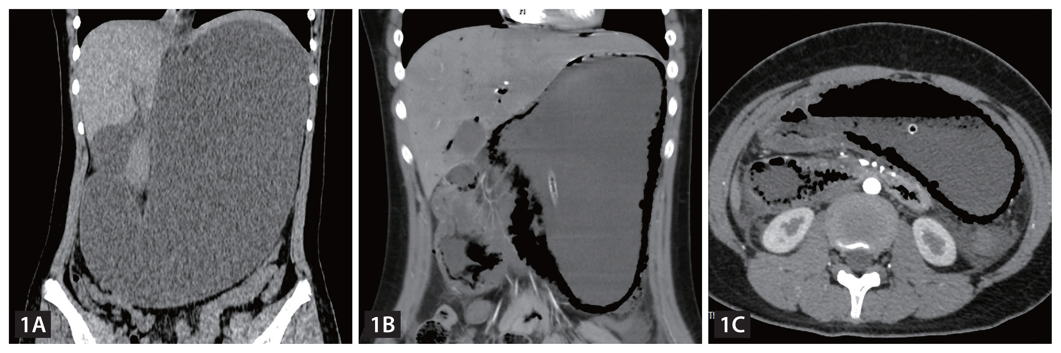
Massive gastric dilatation. (A) Extensive pneumatosis of the gastroduodenal wall and intrahepatic portal vein. (B) The superior mesenteric artery aggravated the degree of duodenal obstruction (C).
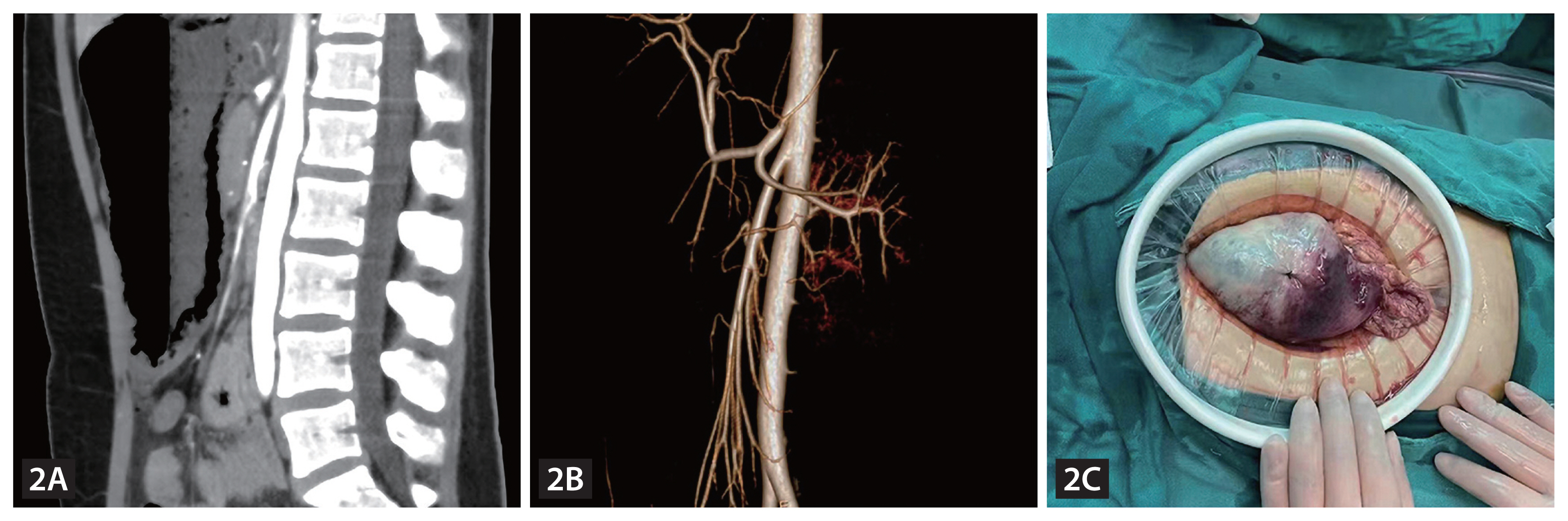
The angle between the superior mesenteric artery and the abdominal aorta was sharply decreased (A, B) Emergency operation revealed that approximately 2/3 of the gastric wall tissue was widely blackened and necrotic (C).
Acute gastric dilatation (AGD) is commonly associated with eating disorders, psychiatric problems, or drug abuse. Superior mesenteric artery syndrome (SMAS) often occurs in patients with chronic decreased body mass index. The possible reasons why the patient had SMAS are as follows: the patient was in a rapidly developing period of adolescence, and the mesenteric fat pad was consumed [1]. Compared with arterial ischemia, venous congestion is more likely to cause gastric necrosis [2]. In this case, SMAS led to slow passage of food masses at the D3 segment of the duodenum and finally induced AGD after accidental overeating. In just ten hours, CT scanning showed that SMAS had promoted the rapid development of simple AGD into extensive gastric wall necrosis and perforation.
In general, a better prognosis can be obtained by treating simple functional AGD through gastrointestinal decompression. For patients with AGD with rapid disease progression and ineffective nonsurgical treatment, we should be alert to the possibility of concurrent SMAS, especially in adolescents.
Acknowledgments
The present study was approved by the Ethics Committee of Affiliated Hospital of Southwest Medical University.
Notes
CRedit authorship contributions
Zhicheng Huang: conceptualization, data curation, formal analysis, writing - original draft; Cao Li: data curation, writing - original draft; Guangcai Tang: writing - review & editing
Conflicts of interest
The authors disclose no conflicts.
Funding
None