Successful coronary angiography via distal radial access in an 82-year-old male patient with polydactyly
Article information

An 82-year-old male patient was admitted due to atypical chest pain, and he underwent coronary angiography (CAG) via a left distal radial access (DRA). We found that he had polydactyly of his left hand, which could be classified as Wassel Type 4 polydactyly (Fig. 1) [1]. We were concerned about the anatomical variation of the left distal radial artery due to the anomaly, but a distal radial pulsation was easily palpated in the anatomical snuff box, and a successful puncture was performed. A CAG was performed using a 5 Fr vascular access sheath 10 cm in length, according to the usual method, and it revealed mild narrowing (Supplementary Video 1-3). We wanted to determine the intact natural vascular anatomy of the polydactyly-affected hand; hence, we performed manual compression for 10 minutes after sheath removal and performed brachial angiography via brachial puncture with a 20G angiocath cannula. Angiography showed the usual run-off of the left distal radial artery through the anatomical snuff box, and there was an additional digital branch to the accessory thumb (Fig. 2, Supplementary Video 4). The puncture site was compressed for an additional two hours with a gauze block and simple elastic bandage to ensure complete hemostasis.
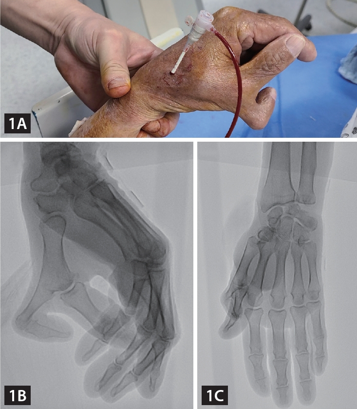
Visual and X-ray images of the patient’s left hand. (A) In the patient’s left hand, an additional proximal phalanx and distal phalanx were observed on the radial side. (B) The X-ray image shows two separate pairs of proximal and distal phalanges articulating at the distal end of the patient’s left 1st metacarpal bone. The additional proximal phalangeal bone was fused with the 1st metacarpal bone. (C) If we ignored the accessory thumb, the overall contour of the left hand appeared normal. Actually, the function of the patient’s left hand was not impaired.

Visual and angiographic images of the patient’s left hand. (A) After removing the guide sheath and applying manual compression for 10 minutes, adequate hemostasis was achieved. (B) Left radial angiography, performed with contrast injection in the left brachial artery, showed that the course of the distal radial artery was not different from that in a normal hand. (C) There was delayed visualization of the ulnar artery, carpal arches, and digital arteries.
Nowadays, in developed countries, most people with polydactyly are treated surgically in their youth; thus, it is rare to find polydactyly persisting in the old age [2]. Therefore, the scenario of a patient with polydactyly undergoing CAG at an advanced age is extremely rare, and this is the first reported case of CAG using DRA in a patient with polydactyly. Written informed consent was obtained from the patient.
Notes
CRedit authorship contributions
Kang Hee Kim: writing - original draft, writing - review & editing, visualization; Bong-Ki Lee: conceptualization, investigation, writing - original draft, writing - review & editing, visualization, supervision, project administration; Jeong Hun Seo: writing - original draft, writing - review & editing, supervision; Kwang Jin Chun: writing - review & editing, supervision; Byung-Ryul Cho: writing - review & editing, supervision
Conflicts of interest
The authors disclose no conflicts.
Funding
None