From pinprick to peril: digital ischemia and necrosis of fingertips induced by routine capillary glucose testing
Article information

A 63-year-old woman with a history of myasthenia gravis, hypertension, and diabetes was referred from Neurology to our department for fingertip lesions evaluation. During her 2-month hospitalization for myasthenia gravis management, she expressed concern about an unexpected lesion on her fingertip that progressively increased in size.
Physical examination revealed no significant lesions on her left hand. However, her right hand’s fingertips displayed variations in skin color from dark brown to black, with advancing necrotic changes, notably on the 5th finger (Fig. 1). Both fingers exhibited pallor from the distal to proximal interphalangeal joint, accompanied by noticeable coldness. Assessment indicated a skin thickness corresponding to a modified Rodnan Skin Score of 2. The patient presented a history of Raynaud’s phenomenon spanning several years. Laboratory analysis revealed an elevated ANA titer of 1:1,280 and anti-centromere antibody level of 63 IU/mL, leading to the definitive systemic sclerosis diagnosis.
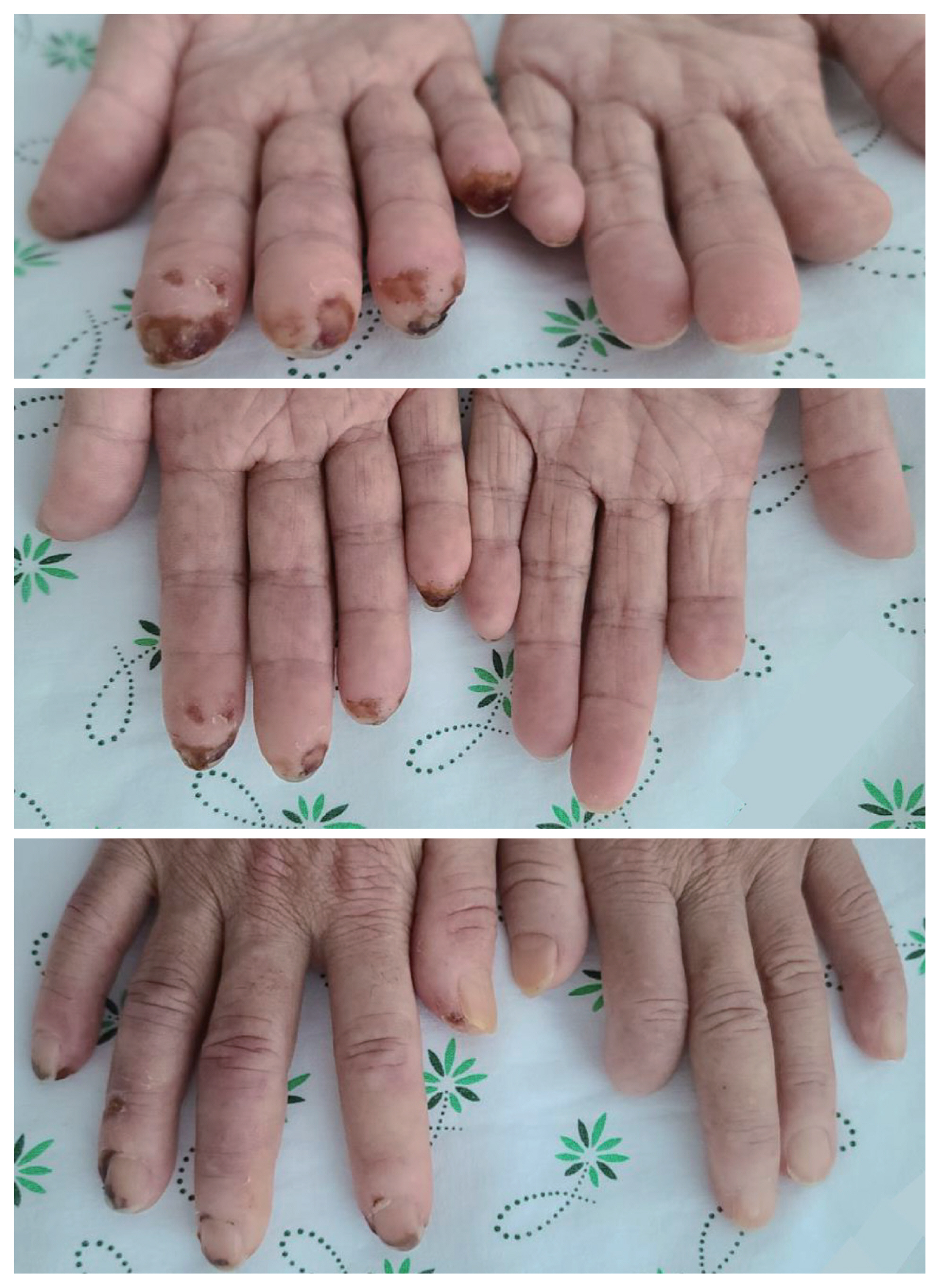
Fingertip ischemia and necrosis in the right hand of a patient with systemic sclerosis following repetitive finger-stick glucose testing. The bilateral fingers exhibited a pallid hue accompanied by the progressive manifestation of cutaneous sclerosis. Particularly noteworthy, the distal phalanges of the right hand—where repetitive capillary glucose testing had been conducted—unveiled conspicuous ischemic transformations, characterized by a spectrum of discoloration spanning from deep brown to ebony black, indicative of incipient necrotic changes.
Despite no recorded trauma, consistent blood glucose testing was conducted on her right hand’s fingers during hospitalization. This suggests that the manifestation of ischemia and necrosis in fingertip regions, particularly where glucose testing was performed, originated from repetitive capillary microinjuries. Despite intravenous prostanoid therapy, progressive necrotic changes in the fifth finger necessitated surgical intervention, leading to the amputation of the distal phalanx.
Systemic sclerosis, an autoimmune connective tissue disease, features fibrosis and vasculopathy. Digital vasculopathy ranges from Raynaud’s phenomenon to ulcers, and necrosis. Mechanical microtrauma alongside ischemia can trigger ulceration [1], causing pain, disability, and tissue loss. Thus, enhancing awareness and prioritizing preventive measures are of utmost importance [2].
Glucose monitoring is pivotal in diabetes management, often involving capillary glucose testing at the fingertips. However, our case study underscores potential complications, including ulceration and necrosis, specifically in systemic sclerosis patients. Consequently, it becomes paramount for clinicians to acknowledge these vulnerabilities among systemic sclerosis patients.
Notes
CRedit authorship contributions
Wan-Hee Yoo: conceptualization, data curation, writing - review & editing, supervision; Yunjung Choi: conceptualization, data curation, writing - original draft, visualization
Conflicts of interest
The authors disclose no conflicts.
The present study was approved by the Institutional Review Board of Chonbuk National University Hospital (CUH 2023-08-065). The study was conducted in accordance with the principles of the Declaration of Helsinki. Informed consent was waived by the board.
Funding
The work was supported by the National Research Foundation of KOREA (NRF) grant funded by the Korea government (MSIT) (No.2021R1G1A1094571).