


INTRODUCTION
Patients who report having diarrhea for more than four weeks should be evaluated for chronic diarrheal illness, since most infectious enteritides and other causes of acute diarrhea generally resolve within this period1–3). Flexible sigmoidoscopy or colonoscopy is often used by many physicians to find the organic cause of chronic diarrhea4), and some gastroenterologists perform routine mucosal biopsies to exclude organic causes of chronic diarrhea that can only be diagnosed microscopically even when the endoscopic findings are normal. Diseases that cause diarrhea when the endoscopic findings are normal or nonspecific include collagenous colitis, lymphocytic colitis and eosinophilic enterocolitis5–7). It is also known that normal-appearing colorectal mucosa in patients with inflammatory bowel disease can show histological abnormalities8–11) and melanosis coli, a sign of laxative abuse, may be seen only microscopically12). Finding these histological abnormalities in grossly normal colonic mucosa may alter the therapeutic decisions by implicating the use of immunosuppressive drugs, such as corticosteroid, or the need for close clinical follow-up. There are controversies surrounding routine mucosal biopsies taken during colonoscopy in patients with normal looking mucosa who have chronic gastrointestinal symptoms. Some suggest colonic biopsies should be taken routinely10, 11, 13), while others argue about the cost-effeciveness of the procedure14, 15). But, it is difficult to conclude the cost-effectiveness of routine mucosal biopsies since these studies had different and obscure inclusion criteria of patients, and different protocols for each biopsy site.
Recently, sporadic cases of these histological abnormalities, especially collagenous colitis, have been reported in Korea16, 17). However, the incidence, of these histological abnormalities among patients with chronic diarrhea has not been reported in Korea.
The purpose of this study is to determine the incidence of clinically important histological abnormalities, prospectively in chronic diarrhea patients with strict inclusion criteria who have grossly normal or nonspecific colonoscopic findings, to ascertain the clinical significance of these findings to therapeutic decision-making and outcome.
MATERIALS AND METHODS
1. Patients
One hundred and eighteen consecutive patients suffering from diarrhea who met our inclusion criteria and visited the gastroenterolgy unit of Samsung Medical Center during a fifteen months period (from April, 1995 to June, 1996) were evaluated prospectively in this study. The inclusion criteria were as follows; 1. patients suffering from chronic nonbloody diarrhea with consistency of watery to loose stool and a frequency of more than 2 times a day for more than a month, 2. no previous history of chronic inflammatory bowel disease, 3. exclusion of patients with specific colonoscopic findings which may explain the cause of diarrhea, such as chronic inflammatory bowel disease, colon cancer and large villous adenoma.
2. Colonoscopic Examinations
Colonoscopic examinations were done with Olympus CF-200I or CF-200L video-colonoscope, and two pieces of biopsies were taken from six different parts of the colon; cecum, ascending colon, mid-transverse colon, descending colon, sigmoid colon and rectum. Of the two biopsy specimens taken from the same part of the colon, one piece was stained with hematoxylin-eosin while the other was stained with Masson-trichrome to determine the thickness of the subepithelial collagen band. Each biopsy specimen was reviewed by a pathologist who had no information on clinical diagnosis prior to interpretation of microscopic findings.
3. Diagnostic Criteria
Collagenous colitis was diagnosed when the subepithelial collagen band thickness, averaging over 10 intercryptal spaces, was measured to 10 μm or greater using the Masson-trichrome stain and signs of epithelial damage, such as epithelial detachments and flattenings, were present. If the subepithelial collagen band thickness was between 5 and 10μm and other features were present, the biopsy was classified as “possible collagenous colitis” Lymphocytic colitis was diagnosed when diffuse presence of intraepithelial lymphocytes was present equal to or more than 10% of colonic epithelial cells and presence of epithelial flattening, increase in crypt lymphocytes, crypt distortion and increase in lamina propria chronic inflammation6). If intraepithelial lymphocyte infiltration was less than 10% but other features of lymphocytic colitis were present, the biopsy was classified as having “some features of lymphocytic colitis”. Eosinophilic enterocolitis was diagnosed when inflammatory cell infiltrate was almost exclusively composed of eosinophils, eosinophilic infiltration outside the GI tract was not present and parasitic infestation was not present16). Melanosis coli was diagnosed if macrophages in the lamina propria displayed cytoplasmic golden pigment. Diagnosis of nonspecific colitis was made when increased lamina propria inflammatory cell infiltrate was present but did not meet the criteria of the above disorders.
4. Biochemical and Microbial Tests
Concurrent microbial, biochemical and physiologic tests were done in a proportion of the patients. These included stool parasite ova, stool occult blood, stool white blood cells, stool fat, stool culture, fasting blood glucose level, serum calcium, phosphorous, thyroid function tests and breath hydrogen test.
RESULTS
Of one hundred and eighteen patients included in this study, 79 were male and the average age was 43.0 years (18–71 years). The average frequency of diarrhea was 3.5±1.7 bowel movements per day and the average duration of symptoms was 45.7±52.4 months (1–240 months).
The gross colonoscopic findings were normal in the majority of cases (71.1%), but some showed nonspecific findings (8.5%), such as mild mucosal edema and hyperemia. Incidental findings, such as small polyps (smaller than 0.7cm, 19 cases), diverticula (3 cases), colonic cyst (1 case), submucosal tumor (1 case) and angiodysplasia (1 case) were discovered during the examinations (Table 1).
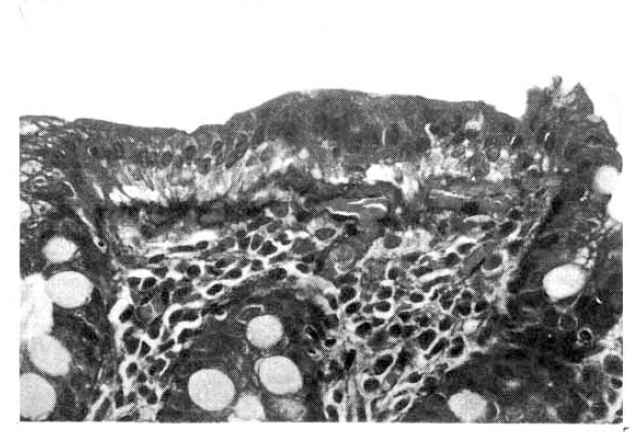
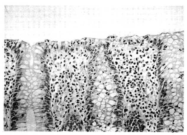
In the microscopic examinations, two cases of collagenous colitis (Fig. 1) and one case of lymphocytic colitis (Fig. 2) and eosinophilic enterocolitis were diagnosed. The gross colonoscopic findings of the cases diagnosed as collagenous colitis and lymphocytic colitis were completely normal, but showed nonspecific edema and hyperemia in the case of eosinophilic enterocolitis. One specimen showed compatible features of ulcerative colitis, but the gross features showed only mild focal mucosal hyperemia at the rectum. Four cases of melanosis coli were discovered during the microscopic examination, gross findings of which were categorized as normal. Sixteen cases (13.5%) of borderline histological abnormalities were observed where 8 cases showed possibility of collagenous colitis and 8 cases showed some features of lymphocytic colitis. Ninety two cases (78.8%) showed nonspecific inflammation only (Table 2).
Results of biochemical, microbial and physiological tests, which were done to a portion of patients, are summarized in Table 3. In particular, fasting hyperglycemia (≥140mg/dl), 2.2% (2/92), hyperthyroidism, 3.8% (2/53) and hypocalcemia, 3.2% (3/93) were discovered. Positive rate of breath hydrogen test was 69.6% (32/46).
In summary, among 118 patients included, 7.6% of patients showed clinically significant histological abnormalities (collagenous colitis, lymphocytic colitis, eosinophilic enterocolitis, ulcerative colitis and melanosis coli), and 13.5% of patients showed borderline histological abnormalities (Fig. 3).
DISCUSSION
Many physicians and gastroenterologists frequently diagnose a patient with chronic diarrhea as irritable bowel syndrome when they get a normal result in colonoscopy, but we had a suspicion that many of those diagnosed as irritable bowel syndrome may have organic causes. Among the various causes of chronic diarrhea, two organic conditions that reportedly cause diarrhea, when colonoscopy and colonic radiography are normal, are collagenous colitis and lymphocytic colitis5–7). Recently, much attention has been paid to those conditions which can be diagnosed by taking biopsies from normal-appearing colonic mucosa, which otherwise would be diagnosed as irritable bowel syndrome. First described in 197619) and 198020), collagenous colitis and lymphocytic colitis respectively have been recognized with increasing frequency in the past few years, as endoscopically normal mucosa has been biopsied more frequently in patients with chronic unexplained diarrhea10, 21–25). Recently, two cases of collagenous colitis have been reported in Korea16, 17), suggesting the possibility that collagenous colitis and lymphocytic colitis might play some etiologic role in chronic diarrhea in Korea.
The two studies recently reported deny the usefulness of a routine mucosal biopsy14–15). Macintosh et al performed sigmoidoscopic biopsies on 134 patients with nonspecific lower gastrointestinal symptoms and reported that they did not find any cases of collagenous colitis or lymphocytic colitis15). Since the symptoms of the majority of patients with collagenous colitis and lymphocytic colitis manifest as chronic watery diarrhea26, 27), they might have included cases that are unlikely to be collagenous colitis or lymphocytic colitis. Only rectal biopsies were performed in their study, which suggest that the cases that are confined to the right side of the colon27) may have been missed. Marshall et al14). have performed mucosal biopsies from normal-looking mucosa obtained from 111 chronic diarrhea patients. They included all patients who experienced a change in their bowel habits, which is a too broad definition of diarrhea. Their data included 1 case of “possible collagenous colitis” and 13 cases that showed some features of lymphocytic colitis of which the clinical significance was denied by the authors. As there have been a few cases that reported development of collagenous colitis in sequential biopsy specimen28), we thought that these cases should not be considered as trivial or nonspecific findings.
Unlike the results by Macintosh, et al.15) and Marshall, et al.14), we were able to find clinically significant histological abnormalities, findings that could alter therapeutic decisions in 7.6% of patients (9/118); 2 cases of collagenous colitis and 1 case for each of the following-lymphocytic colitis, eosinophilic enterocolitis, ulcerartive colitis and 4 cases of melanosis coli. The differences between our result and the previous reports can be accredited to more strict patient selection, more broadened biopsy field and possibly a racial difference between Caucasians and Asians. We also think that the borderline histological abnormality group (13.5%) observed in our study should be followed carefully for a possible development of collagnous colitis or lymphocytic colitis. Careful long-term clinical follow-up and, if necessary, follow-up colonoscopy with biopsies are mandatory.
It should be noted that when collagenous colitis or lymphocytic colitis is suspected, biopsy sites should include the whole length of the colon, including the right side colon. The lesions can be discontinuous and thus absent in some biopy specimens, even in multiple specimens from a given area29). It is difficult to determine the optimal number of biopsies that should be taken, and from what locations within the colon but, based on available information, we suggest that, at least six, biopsies should be taken approximately evenly spaced between the cecum and the rectum to exclude those conditions.
Our results of the biochemical and microbial tests need to be mentioned. Although our results cannot be used to define the relative etiologic roles of each metabolic and biochemical cause of chronic diarrhea, since the tests were not performed in all of the patients included in the study, they provide valuable information that metabolic causes, such as diabetes melitus, hyperthyroidism, hypocalcemia and microbial causes, such as parasitic infestation, may play a role in a significant proportion of the patients. Our results show that 69.6% of patients (32/46) were positive for breath hydrogen test, but it was not possible to attribute the cause of the patients’ symptoms to lactose intolerance because therapeutic trials of lactose free diet were not performed on those who were positive for breath hydrogen test. It is peculiar to find that the positive rate of breath hydrogen breath test showed lower than the expected positive rate in general Korean patients, which is approximately 85%30). We speculate that this low rate of positive breath hydrogen could be due to false negative tests, which may reflect increased small bowel motility in patients with chronic diarrhea.
In conclusion, clinically significant histological abnormalities can exist in significant percentages, in spite of normal or nonspecific colonoscopic findings, which can justify routine mucosal biopsy in the evaluation of chronic diarrhea patients. The clinical significance of borderline histological abnormalities needs to be determined by careful follow-up studies.

 PDF Links
 PubReader
 ePub Link
 Full text via DOI
 Download Citation
 Print
Print


