Korean J Intern Med > Volume 2(1); 1987 > Article
Chung, Lee, Park, Sohn, Park, and Shin: Ferritin Assay in Malignant Pleural Effusion

Abstract

In order to assess the usefulness of ferritin as a tumor marker, the authors measured and analyzed pleural fluid and serum ferritin concentrations by double antibody radioimmunoassay method in 20 patients with malignant pleural effusion, and in 39 patients with benign pleural effusion. Serum ferritin levels were also measured in a control group of 20 healthy people.
The results obtained are summarized as follows:
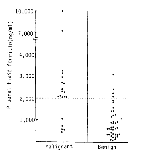
  • 1) Pleural fluid ferritin levels in the malignant pleural effusion group were significantly higher (p<0.001) than those of the benign pleural effusion group.

  • 2) As one of the criteria in differentiating between malignant and benign pleural effusion, the differentiating pleural fluid ferritin level was set at 2,000 ng/ml, a specificity up to 75.0% and sensitivity of 89.7% could be obtained.

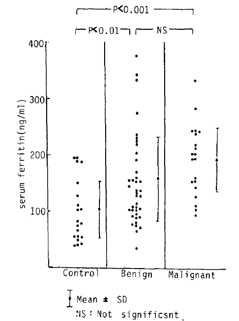
  • 3) Serum ferritin levels in the malignant pleural effusion group were significantly higher (p<0.001) than those in the control group.

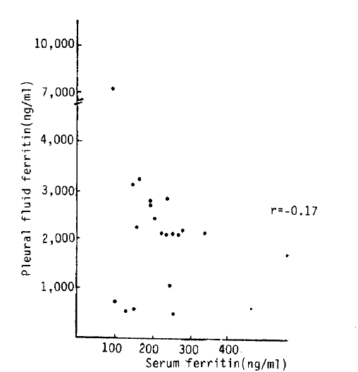
  • 4) There was no statistically significant correlation between pleural fluid and serum ferritin levels in the malignant pleural effusion group.

From the above results, it can be concluded that it is possible to use pleural fluid ferritin levels as a tumor marker.

INTRODUCTION

Ferritin, an iron containing, iron storage protein, exists in the human liver, spleen and bone marrow. Ferritin levels closely reflect the amount of iron in the body15), and the serum ferritin test is known to be the most accurate mothod for the assessment of the amount of body iron stores. Moreover, serum ferritin is known to be increased not only in various liver diseases and inflammatory diseases but also with malignant tumors1,4,612), and there have been attempts to use ferritin as a tumor marker1).
Differential diagnosis of pleural effusion is one of the most frequent difficulties in the field of pulmonology. A significant portion of pleural effusions are caused by malignant tumors13) and differentiation those caused by benign tumors just by thoracentesis and clinical manifestation is often difficult and usually requires cytologic or histologic examination as well. Malignant pleural effusion can be diagnosed in about 60% of patients by cytologic examination and in about 40–60% by pleural biopsy. When both of the above two methods are performed, diagnosis can be confirmed in up to 60–90% of cases14). Even when both cytologic and histologic examinations are carried out together, the differential diagnosis still remains difficult in some cases. It has been reported that measurement of orosomucoid, β2-microglobulin or carcinoembryonic antigen (CEA) in the pleural fluid can be used as an additional examination to assist in the differential diagnosis of pleural effusion1518).
This is a report of ferritin assays in both serum and pleural fluid in patients with pleural effusions to see whether ferritin could be used as a reliable marker to differentiate between benign and malignant causes or not. The results were analyzed in regard to diagnostic significance and use as criteria for differential diagnosis.

SUBJECTS AND METHODS

1. Subjects

The study was done on 59 patients who were admitted to Pusan National University Hospital between February 1985 and August 1986 because of pleural effusion. All had undergone extensive investigations in order to diagnose the causes of the effusion. Among them, there were 20 cases with malignant pleural effusion confirmed by case history, clinical symptoms, chest roentgenography and computerized tomography, cytologic examination of sputum and pleural exudate, pleural biopsy, lymph node biopsy and percutaneous transthoracic needle aspiration and biopsy. There were 39 cases of benign pleural effusion confirmed by pleural biopsy, and smear and culture of sputum for tubercule bacilli and which also included cases that showed an improvement on antituberculosis chemotherapy.
The control group consisted of 20 healthy subjects, 10 of each sex. Among the 20 cases of malignant pleural effusion, the most common type was primary bronchogenic carcinoma with 14 cases; 6 adenocarcinoma, 5 squamous cell carcinoma, 2 small cell carcinoma, and 1 mucoepidermoid tumor. The remainder were metastatic carcinoma patients, 2 of known origin, namely 1 case each from breast and stomach and 4 of unknown origin, namely, 3 cases of adenocarcinoma and 1 case of undifferentiated carcinoma.
Among the 39 cases of benign pleural effusion, there were 26 with tuberculous pleurisy, 3 with liver cirrhosis with ascites, 3 with nephrotic syndrome, 2 with chronic renal failure, 2 with congestive heart failure, and 1 case each of pneumonia, empyema, and interstitial lung disease.

2. Methods

The measurements of the pleural fluid and serum ferritin concentrations were made by using a double antibody radiolabeled 125I-ferritin radioimmunoassay kit produced by Diagnostic Products Corporation of U.S.A.. When the concentration exceeded the level of 1,000 ng/ml, dilution measurements were used instead, reading up to 10,000 ng/ml.

RESULTS

1) There was a significant difference in mean pleural fluid ferritin levels between benign and malignant pleural effusions with levels of 894.4±729.9 ng/ml and 2656±2194,2 ng/ml respectively (p<0.001) (Table 3, Fig. 1 and 2).
When the mean serum ferritin levels were compared between the 2 groups of pleural effusion, there was no significant difference, benign effusion 160.8±80.6 ng/ml and malignant effusion 193.3±60.2 ng/ml, but when these levels were compared separately to the mean serum ferritin level of the control group, 103.9±56.0 ng/ml a significant difference was seen, p<0.01 with benign effusion and p<0.001 with malignant effusion (Table 4 and Fig. 2).
2) In the malignant pleural effusion group pleural fluid ferritin levels in the 14 cases of primary bronchogenic carcinoma were compared to levels in 6 cases of metastatic cancer. There was no difference between the two groups, being 3105. 8±2379.5 ng/ml in primary bronchogenic carcinoma and 1554.8±1075.8 ng/ml metastatic cancer.
There was also no difference in serum ferritin levels, being 192.4±54.7ng/ml in primary bronchogenic carcinoma and 195.2±71.4 ng/ml in metastatic cancer (Table 5).
3) In the malignant pleural effusion group the pleural fluid and serum ferritin levels were compared between 10 cases of adenocarcinoma and 10 cases of non-adenocarcinoma. The mean pleural fluid ferritin level in adenocarcinoma group was 2491.1±1748.0 ng/ml and in non-adenocarcinoma group was 2789.9±2555.7 ng/ml. The mean serum ferritin level in adenocarcinoma was 181.4±55.3 ng/ml and in non-adenocarcinoma was 205.1±62.5 ng/ml. No statistically significant difference was present between the 2 groups in either study (Table 6).
4) There was no statistically significant correlation between mean pleural fluid ferritin and serum ferritin levels in the malignant pleural effusion group (r= −0.17) (Fig. 3).
5) The distribution of ferritin levels in pleural fluid was studied. In the malignant pleural effusion group, the minimum level was 498 ng/ml and the maximum level 10,000 ng/ml. There were 4 cases (20.0%) ranging between 101–1,000 ng/ml, 1 case (5.0%) between range of 1,001–2,000 ng/ml, 13 cases (65.0%) between the range of 2,001–5,000 ng/ml, and 2 cases (10.0%) between the range of 5,001–10,000 ng/ml. In the benign pleural effusion group, the range was between a minimum level of 56 ng/ml and a maximum level of 3014 ng/ml. There were 3 cases (7.7%) between the range of 1–100 ng/ml, 24 cases (61.5%) between the range of 101–1,000 ng/ml, 8 cases (20.5%) between the range of 1,001–2,000 ng/ml and 4 cases (10.3%) between the range of 2,001–5,000 ng/ml (Table 7 and Fig. 1).
6) According to these results when a pleural fluid ferritin level of 2,000 ng/ml was taken as the level to differentiate between malignant and benign effusions the ferritin level was above this point in 15 out of the 20 cases of malignant effusion, showing a sensitivity of 75.0% and specificity of 89.7%.

DISCUSSION

Ferritin was first separated from the liver and spleen of the horse by Laufberger in 193719). Ferritin is one of the most essential iron storage proteins and can be detected in most tissues but is mainly concentrated in liver, spleen, and bone marrow. It forms 15–20% of total body iron content20,21).
Apoferritin which surrounds the outer wall of the ferritin molecule, is synthesized mainly from free polysome of the liver and partially from the endoplasmic reticulum22). Iron injected into the body stimulates the synthesis of apoferritin and is transported by transferrin to be utilized in the formation of iron micelle in a form of Fe++. To this iron micelle 24 apoferritins bond together and form a globular shaped ferritin23,24). 5,000 ions of iron bond to one ferritin molecule and the molecular weight of the ferritin molecule is 450,0001).
Ferritin consists of many different kinds of isoferritin, but may be divided broadly into two categories of basic isoferritin and acidic isoferritin according to it’s isoelectric point, since each tissue ferritin has a different motility under electrophoresis and isoelectric focusing2528). It has been shown that basic isoferritin exists mainly in the liver and spleen and forms most of the serum ferritin, whereas acidic isoferritin is known to be present in heart, kidney, pancreas and placenta20,2931) A rjse of the serurn ferritin level6,1012) and changes in its composition32,33) in various malignant tumors have been reported, and since both tissue and serum ferritin in malignant tumors are known to be acidic isoferritin, there have been attempts to use acidic isoferritin as a tumor marker11,31,33). Maxim et al.34) reported an increase in ferritin levels in lung cancer tissue. Serum ferritin, together with carcino-embryonic antigen (CEA), has been reported to be useful in indicating tumor recurrence and thus in postoperative follow up35,36).
In this experiment there was no statistically significant difference between serum ferritin levels of the malignant pleural effusion group of 193.3±60.2 ng/ml and the benign effusion group of 160. 8±80.6 ng/ml. However when the levels of each group were compared separately to the serum ferritin levels of the control group, 103.9±56.0 ng/ml, both groups showed a statistically significant difference, p<0.001 and p<0.01 respectively. These results are compatible with other reports that serum ferritin levels increase both in various inflammatory diseases and in malignant tumors6,912).
As the pleural fluid ferritin levels in malignant and benign effusions showed a statistically significant difference, 2656.4±2194.2 ng/ml and 894.2±729.9 ng/ml respectively (p<0.001), the possibility that pleural fluid ferritin measurement could be used as a tumor marker to assist in the differentiation between malignant and benign pleural effusion was investigated.
If the differentiating level for pleural fluid ferritin was set at 2,000 ng/ml, the ferritin level was positive in 15 out of the 20 cases of malignant pleural effusion indicating a sensitivity of 75.0% and specificity of 89.7%. When the above result was compared to the reports of Millano37) and Jung et al.38) their differentiating levels were similar, at 2,000 ng/ml and 1,900 ng/ml respectively. However, their results of sensitivity were only 34% and 69% respectively, much lower than the results of this study. The authors are of the opinion that the disparity in sensitivity could be due to the difference in the subjects studies and to the methods used in measuring ferritin levels.
When pleural fluid CEA, was used as a tumor marker, James et al.39) reported that high levels of pleural fluid CEA were present when the cause of the pleural effusion was adenocarcinoma.
However, in this experiment, there was no significant difference in levels of pleural fluid ferritin between adenocarcinoma and non-adenocarcinoma group. Moreover, there was also no statistically significant difference between primary lung cancer and metastatic cancer groups.
The possible causes of the rise in ferritin levels in malignant pleural effusions can be listed as follows: first, an influx of serum ferritin into the plerual fluid, secondly, hemolysis of erythrocytes and thus release of ferritin from erythrocytes, and most of the malignant pleural effusions are bloody, and thirdly, as malignant tumor invades the pleura, increased synthesis of ferritin from malignant tumor cells and an influx of ferritin from the surrounding inflammatory tissues occurs38).
The authors only measured plerual fluid and serum ferritin levels in order to make a differential diagnosis of malignant pleural effusion, however, if the already widely used CEA as a tumor marker17,18) had been measured at the same time, probably, results of a higher sensitivity and specificity would have been obtained.

Fig. 1.
Levels of pleural fluid ferritin in patients with malignant and benign pleural effusion.
kjim-2-1-106-18f1.gif
Fig. 2.
Comparison of serum ferritin levels in normal control group & patients with benign & malignant pleural effusion.
kjim-2-1-106-18f2.gif
Fig. 3.
Relationship between pleural fluid & serum ferritin in patients with malignant pleural effusion.
kjim-2-1-106-18f3.gif
Table 1.
Etiologies of Malignant Pleural Effusion
Diagnosis No. of patients
Lung cancer 14
  Adenocarcinoma 6
  Squamous carcinoma 5
  Small cell carcinoma 2
  Mucoepidermoid tumor 1
Metastatic cancer 6
  Breast (ductal cell carcinoma) 1
  Stomach (adenocarcinoma) 1
  Unknown origin
    Adenocarcinoma 3
    Undifferentiated carcinoma 1

Total 20
Table 2.
Etiologies of Benign Pleural Effusion
Diagnosis No. of patients
Tuberculous 26
Liver cirrhosis 3
Nephrotic syndrome 3
Chronic renal failure 2
Congestive heart failure 2
Pneumonia 1
Empyema 1
Interstitial lung disease 1

Total 39
Table 3.
The Mean Values of Pleural Fluid & Serum Ferritin in Benign & Malignant Pleural Effusion
Group Pleural fluid (ng/ml) Serum (ng/ml)
Benign effusion 894.2±729.9 160.8±80.6
Malignant effusion 2,656.4±2,194.2 193.3±60.2

P value P < 0.001 NS

Values are expressed as mean ±SD.

NS: Not significant

Table 4.
The Mean Values of Serum Ferritin in Control Group, Benign & Malignant Pleural Effusion
Group Serum ferritin (ng/ml) P value
Control 103.9±56.0
Benign effusion 160.8±80.6 P < 0.01
Malignant effusion 193.8±60.2 P < 0.001

Values are expressed as mean ±SD.

Table 5.
The Mean Values of Pleural Fluid & Serum Ferritin in Primary & Metastatic Lung Cancer
Group Pleural fluid (ng/ml) Serum (ng/ml)
Primary 3,105.8±2,379.5 192.4±54.7
Metastatic 1,554.8±1,075.8 195.2±71.4

P value NS Ns

Values are expressed as mean ± SD.

NS: Not significant

Table 6.
The Mean Values of Pleural Fluid & Serum Ferritin in Adenocarcinoma & Non-adenocarcinoma
Group Pleural fluid (ng/ml) Serum (ng/ml)
Adenocarcinoma 2,491.1±1,748.0 181.4±55.3
Non-adenocarcinoma 2,789.9±2,555.7 205.1±62.5

P value NS NS

Values are expressed as mean ±SD.

Table 7.
Distribution of Pleural Fluid Ferritin in Patients with Malignant & Benign Effusion
Pleural fluid ferritin (ng/ml) No. of patients with malignant effusion (%) No. of patients with benign effusion (%)
1 – 100 3 (7.7)
101 – 1,000 4 (20.0) 24 (61.5)
1,001 – 2,000 1 (5.0) 8 (20.5)
2,001 – 5,000 13 (65.0) 4 (10.3)
5,001 – 10,000 2 (10.0)

Total 20 (100.0) 39 (100.0)

REFERENCES

1. Jacobs A, Worwood M. Ferritin in serum: clinical and biochemical implications. N Engl J Med 292:951. 1975.
crossref pmid
2. Jacobs A, Miller F, Worwood M, Beamish MR, Wardrop CA. Ferritin in the serum of normal subjects and patients with iron deficiency and iron overload. Br med J 28:206. 1972.
crossref
3. Jacobs A. Serum ferritin and iron stores. Federation procedings 36:2024. 1977.

4. Lipschitz DA, Cook JD, Finch CA. A clinical evaluation of serum ferritin as an index or iron stores. N Engl J Med 290:1213. 1974.
crossref pmid
5. Kim BK, Chang YB, Cho BY, Lee HK, Koh CS, Lee MH. Studies on the serum ferritin assay as an evaluation of storage iron. Kor J Intern Med 24:2. 1982.

6. Rew JS, Kahng HK, Chang KS, Joh NJ, Yoon CH. Studies on the serum ferritin in various malignant tumors. Kor J Intern Med 25:7. 1982.

7. Lee WP, Kim SK, Park SB, Yoon YB, Kim CH, Im JS. Serum ferritin levels in the patients liver diseases. Kor J Gastroenterol 14:1. 1982.

8. Prieto J, Barry M, Sherlock S. Serum ferritin in patients with iron overload acute and chronic liver diseases. Gastroenterology 68:525. 1975.
crossref pmid
9. Birgegard G, Hallgren R, Killander A, Stromberg A, Venge P, Wide L. Serum Ferritin during infection: A longitudinal study. Scand J Haematol 21:333. 1978.
crossref pmid
10. Marcus DM, Zimberg N. Isolation of ferritin from human mammary and pancreatic carcinomas by means of antibody adsorbents. Ach Biochem Biophysiol 162:493. 1974.
crossref
11. Alpert E. Characterization and subunit analysis of ferritin isolated from normal and malignant human liver. Cancer Res 35:1505. 1975.
pmid
12. While GP, Worwood M, Parry DH, Jacobs A. Ferritin synthesis in normal and leukemic leukocyte. Nature 250:584. 1974.
crossref pmid
13. Hirsch A, Ruffie P, Nebut M, Bignon J, Chretien J. Pleural effusion: Laboratory tests in 300 cases. Thorax 34:106. 1979.
crossref pmid pmc
14. Salyer WR, Eggleston JC, Ernzan YS. Efficacy of pleural needle biopsy and pleural fluid cytopathology in the diagnosis of malignant neoplasm involving the pleura. Chest 670:536. 1975.
crossref
15. Agostoni A, Marasini B. Orosomucoid contents of pleural and peritoneal effusions of various etiologies. Am J Clin pathol 67:146. 1977.
crossref pmid
16. Vladutiu AO. β2-microglobulin in pleural fluids. N Engl J Med 294:903. 1976.
crossref
17. Rittgers RA. Carcinoembryonic antigen levels in benign and malignant pleural effusions. Ann Intern Med 88:631. 1978.
crossref pmid
18. Jang YS, Jang SH, Sohn HY, Kim SK, Lee WY, Kim KH. A study for carcinoembryonic antigen and ratios of pleural fluid to serum immunoglobulins in pleural effusion. Kor J Intern Med 29:50. 1985.

19. Laufberger V. Sur la cristallisation de la ferritine. Bull Soc Chem Biol 19:1575. 1937.

20. Munro M, Linder H. The function and distribution of ferritin. Physiol Rev 58:317. 1978.
crossref pmid
21. Robert R, Crichton B. Ferritin: structure, synthesis and function. N Engl J Med 284:1413. 1971.
crossref pmid
22. Puro DG, Richter GW. Ferritin synthesis by free and membrane (poly) ribosome of rat liver. Proc Soc Exp Biol Med 138:399. 1971.
crossref pmid
23. Harison PM, Hoarse RJ, Hoy TG. Ferritin and hemosiderin structure and function, Iron in biochemistry and medicine. London: Academic Press, 73. 1974.

24. Lones T, Spencer R, Walsh C. Mechanism and kinetics of iron release from ferritin by dihydroflavins. Am Chem 17:4011. 1978.

25. Linder-Horwitz M, Ruettinger RT, Munro HN. Iron induction of electrophoretically-different ferritins in rat liver, heart, and kidney. Biochem Biophysiol Acta 200:442. 1970.
crossref
26. Gabuzda TG, Pearson J, Melvin M. Metabolic and molecular heterogeneity of marrow ferritin. Blood 29:770. 1967.
crossref pmid
27. Crichton RR, Miller JA, Cumming RLC. The organ specificity of ferritin in human and horse liver and spleen. Biochem J 131:51. 1974.
crossref
28. Massover WH. Multiple isoferritin in mouse liver: Demonstration by polyacrylamide gel electrophoresis. Biochem Biophysiol Acta 532:202. 1978.
crossref
29. Drysdale JW, Singer RM. Carcinofetal human isoferritins in placenta and HeLa cells. Cancer Res 34:3353. 1974.

30. Drysdale JW, Adelman TG. Human isoferritin in normal and disease states. Semin Hematol 14:71. 1977.
pmid
31. Powell LW, Alpert E, Isselpacher KJ, Drysdale JW. Human isoferritins: Organ specific iron and apoferritin distribution. Br J Hematol 30:47. 1975.
crossref
32. Makino Y, Konno K. A comparison of ferritin from normal and tumor bearing animals. J Biochem 65:471. 1969.
crossref pmid
33. Alpert E, Coston RL, Drysdale JW. Carcinofetal human liver ferritin. Nature 242:194. 1973.
crossref pmid
34. Maxim PE, Prather JR, Veltri RW. Ferritin levels in tissue extracts and serum of patients with carcinoma of the lung. Fed Proc 39:413. 1980.

35. Vincent RG. Biologic markers in lung cancer. Seminars in Res Med 3:184. 1982.
crossref
36. Gropp C, Hanemann K. Carcinoembryonic antigen and ferritin in patients with lung cancer before and during therapy. Cancer 42:2802. 1978.
crossref pmid
37. Milano C. Use of tumor marker as a supplement to cytology in diagnosis of malignant effusions. Clinical Chemistry 26:1632. 1980.
crossref
38. Chung YT, Han YC. Ferritin assay in plerural effusion. Tuberculosis and Respiratory 31:10. 1984.

39. James Mckenna A, Chandrasekhar AJ, Henkin RE. Diagnostic value of carcinoembryonic antigen in exudative pleural effusion. Chest 78:587. 1980.
crossref pmid

Editorial Office
101-2501, Lotte Castle President, 109 Mapo-daero, Mapo-gu, Seoul 04146, Korea
Tel: +82-2-2271-6792    Fax: +82-2-790-0993    E-mail: kaim@kams.or.kr                

Copyright © 2024 by Korean Association of Internal Medicine.

Close layer
prev next