


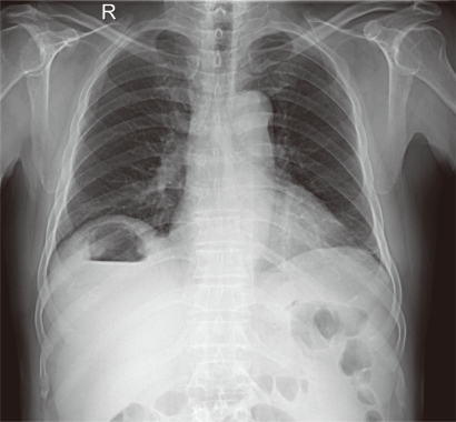
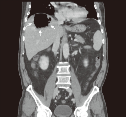
A 70-year-old man with a history of diabetes mellitus presented with weakness and poor oral intake for 1 month. On physical examination, he was febrile and had mild right upper quadrant tenderness. The patient's white cell count was 14,900/mm3, with 79.0% neutrophils. A chest radiograph showed an air-fluid level in the right-upper abdomen (Fig. 1). Computed tomography (CT) revealed a huge gas-forming liver abscess (Fig. 2). The patient underwent sonography-guided drainage, and was given intravenous antibiotics and insulin to control his diabetes. The culture of the pus grew Klebsiella pneumoniae. He recovered and was discharged.
The diagnostic tools for gas-forming pyogenic liver abscess (GPLA) are sonography and CT. On plain films, an air-fluid level or mottled gas pattern is the most common finding. Gas within the liver parenchyma is reported to be visible in up to 36% of patients with GPLA. Unless suspected, this may be mistaken for bowel gas. In our case, we saw an air-fluid level in the right upper abdomen in the plain X-ray, which enabled us to detect GPLA early.
 PDF Links
 PubReader
 ePub Link
 Full text via DOI
 Download Citation
 Print
Print


