


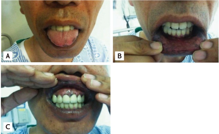
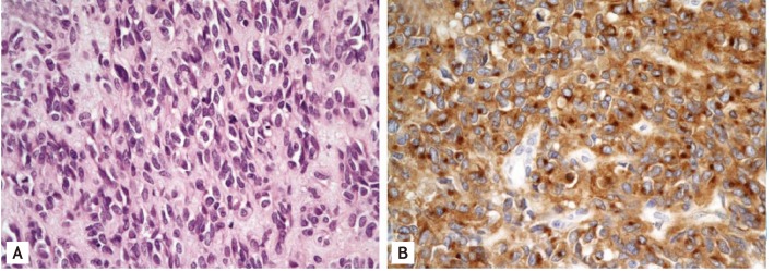
A 45-year-old male presented with a 3-week history of hyperpigmentation of the lip, tongue, and oral cavity (Fig. 1). He had attended an endocrinology outpatient clinic for evaluation of diabetes or adrenal insufficiency. The patient did not have diabetes, but had a 7-year history of hypertension. The endocrinologist diagnosed acanthosis nigricans clinically. On systematic review, the patient stated that he had abdominal distension, discomfort, and easy satiety for several months. He was referred to us for an evaluation of the abdominal discomfort and internal malignancy. Computed tomography of the abdomen and pelvis revealed an enhancing infiltrative mass involving the entire peritoneum in the mid to lower and left upper quadrants of the abdomen and a small amount of ascites in the perihepatic space extending into the right paracolic gutter and pelvic cavity. This suggested a peritoneal carcinomatosis-like malignancy involving the peritoneum, such as peritoneal mesothelioma diffuse type, lymphoma, or desmoplastic small round cell tumor (Fig. 2A). A peritoneoscopic biopsy showed diffuse scattered nodules up to 1 cm on the viscer omentum (Fig. 2B). These bled readily on touch. Histological and immunohistochemical analyses of the biopsy specimen showed diffusely c-kit-positive epithelioid cells, which are consistent with a malignant gastrointestinal stromal tumor (GIST) (Fig. 3). After diagnosing a GIST, we planned palliative chemotherapy with imatinib as it was inoperable. However, the patient was lost to follow-up.
Acanthosis nigricans is a paraneoplastic dermatosis characterized by velvety, hyperpigmented plaques on the skin in body folds, typically the armpits, groin, and neck. Sometimes the lips, palms, or soles are affected. Malignant acanthosis nigricans is associated with various neoplasms, most often gastrointestinal adenocarcinoma. The association of this condition with sarcoma is extremely rare. Here, we report acanthosis nigricans on the lip and oral mucous membrane in a patient with GIST. To our knowledge, this is the first case of acanthosis nigricans associated with a malignant GIST.

 PDF Links
 PubReader
 ePub Link
 Full text via DOI
 Download Citation
 Print
Print


