


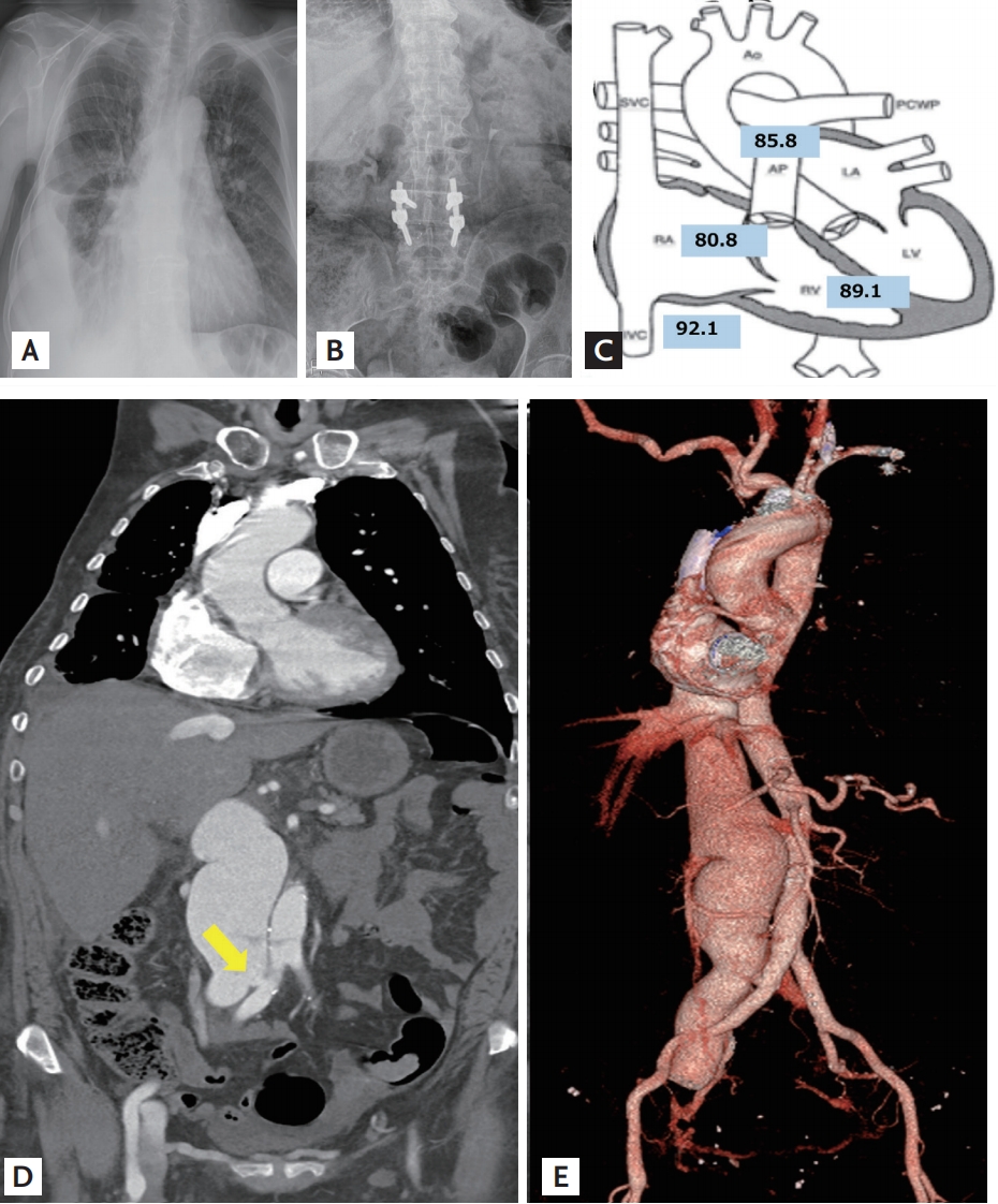
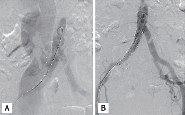
A 62-year-old woman was referred to our hospital with recurrent right pleural effusion after lumbar spine surgery 18 months earlier. Immediately after the surgery, she received medical treatment for pneumonia and right heart failure (Fig. 1A and Fig. 1B). Her symptoms persisted despite tricuspid valve annuloplasty for tricuspid regurgitation. She received thoracentesis (1 to 2 L per week). An echocardiogram showed a dilated right heart, increased pulmonary arterial pressure (maximum pressure gradient: 59 mmHg) and normal ejection fraction. At first, we suspected chronic thromboembolic pulmonary hypertension and proceeded with the computed tomography (CT) pulmonary angiogram, but results were negative. To identify the cause of pulmonary hypertension, we performed right heart catheterization. Pulmonary capillary wedge pressure was 23 mmHg and the ratio of pulmonaryŌĆösystemic blood flow was 2.66, indicating the presence of left to right shunt. The highest oxygen step-up was confirmed in the inferior vena cava (IVC) (Fig. 1C). Transesophageal echocardiography and heart magnetic resonance were negative for intracardiac shunt. During auscultation of lung sounds 12 days post-admission, we found a holosystolic murmur in her back. She underwent aorta CT angiography for detection of an extracardiac shunt. An arteriovenous fistula between the right common iliac artery and IVC was found (Fig. 1D and Fig. 1E). A 16 ├Ś 82 mm sized stent graft was deployed across the fistula (Fig. 2). After the procedure, her symptoms and right pleural effusion did not relapse.
In our case, arteriovenous fistulas may be formed immediately when the laceration includes both arterial and venous tearing during lumbar surgery. Multiple tests and time were needed to find the cause of right heart failure. Had we detected her abnormal lung sounds earlier, the diagnosis would have been faster, without unnecessary tests. This case demonstrates that the integration of clinical history and physical exam results in differential diagnosis is crucial for timely diagnosis.
Written informed consent was obtained from the patient who participated in this study.
 PDF Links
 PubReader
 ePub Link
 Full text via DOI
 Download Citation
 Print
Print


