Korean J Intern Med > Volume 40(6); 2025 > Article
Ohn, Choi, Yoo, Seo, and Lee: Left main coronary artery compression associated with unfavorable thoracic anatomy
A 23-year-old male presented with exertional chest pain that began a month prior. He had undergone patent ductus arteriosus closure in the neonatal period and exhibited no significant findings on physical examination except for a shallow chest cavity.
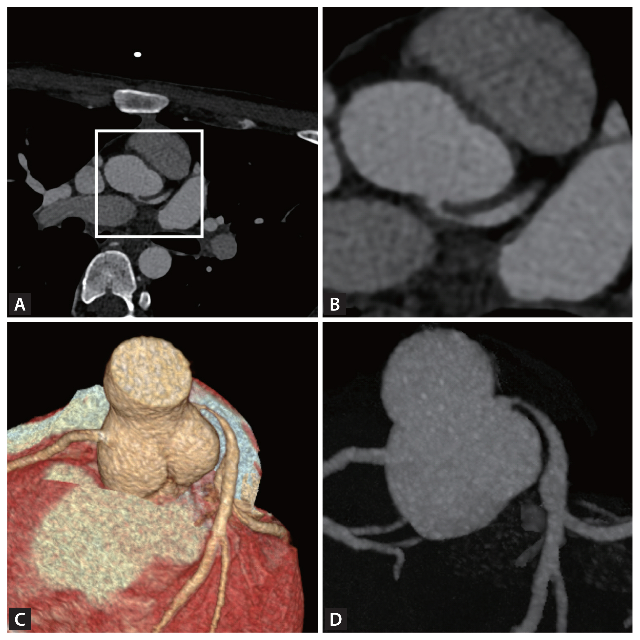
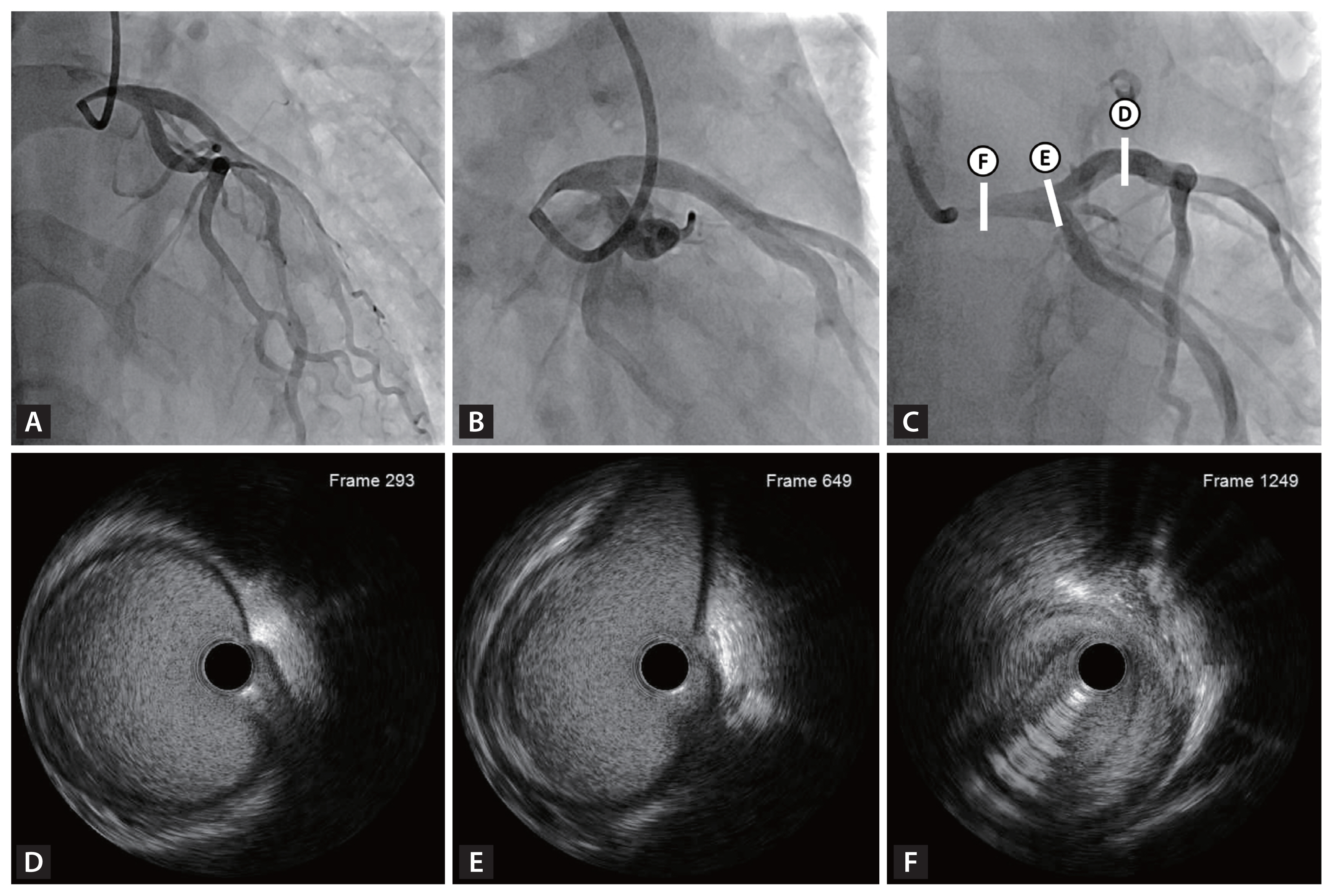
Electrocardiography and echocardiography results were within normal limits. However, cardiac computed tomography angiography revealed significant left main coronary artery (LMCA) stenosis due to an acute angle and relatively high takeoff of the LMCA, coursing between the aorta and pulmonary trunk (Fig. 1, Supplementary Video 1). Subsequent coronary angiography confirmed significant LMCA stenosis, and intravascular ultrasound demonstrated gradual narrowing of the proximal vessel lumen compared to distal reference, suggesting extrinsic compression rather than plaque accumulation. (Fig. 2). Considering the patient’s overall clinical assessment and multimodal imaging findings, he underwent coronary artery bypass graft surgery and recovered uneventfully.
Coronary compression due to unfavorable anatomy has been associated with anomalous aortic origin of a coronary artery [13]. While uncommon, this condition can manifest as exertional chest pain in adolescence and can lead to coronary compromise, including sudden cardiac death [13]. Although this case does not fit within the category of congenital anomaly, it shares anatomical characteristics affecting the takeoff and course of the LMCA. Furthermore, LMCA compression, though extremely rare, is clinically significant. Therefore, in young patients with exertional chest pain, early imaging is crucial to distinguish extrinsic vascular compression from primary coronary artery disease and to guide appropriate management.
Informed consent was obtained from patients.

Supplementary Information

Notes

CRedit authorship contributions
Daeung Ohn: conceptualization, investigation, writing - original draft, writing - review & editing, visualization; Yeon-Jik Choi: writing - review & editing, project administration; Jin-Young Yoo: writing - review & editing, visualization; Suk-Min Seo: writing - review & editing, supervision; Junghoon Lee: writing - original draft, writing - review & editing, supervision, project administration
Conflicts of interest
The authors disclose no conflicts.
Funding
None

Figure 1
Cardiac computed tomography angiography of the left coronary artery. (A) An axial image of cardiac computed tomography angiography demonstrates severe stenosis at the ostium of the left main coronary artery, which exhibits a high take-off with an acute angle and an interarterial course. (B) Enlarged view of the origin of the left main coronary artery, corresponding to the boxed area in (A). (C, D) Three-dimensional volume-rendered and maximum intensity projection images illustrate a relatively high take-off with an acute angle of the left main coronary artery.
kjim-2025-172f1.jpg
Figure 2
Coronary angiography and corresponding intravascular ultrasound (IVUS) images of the left coronary artery. (A, B) Coronary angiography revealed significant focal stenosis in the proximal left main artery, while the distal branches demonstrated preserved flow. (C) Coronary angiography with markers indicating the locations of IVUS cross-sectional images. (D, E) IVUS frames of the proximal left anterior descending artery and distal left main coronary artery show normal vessel anatomy without atherosclerosis. (F) IVUS frame of the proximal left main coronary artery demonstrates a luminal narrowing without significant plaque burden, likely due to external compression caused from anatomical deformity.
kjim-2025-172f2.jpg

REFERENCES

1. Cheitlin MD, De Castro CM, McAllister HA. Sudden death as a complication of anomalous left coronary origin from the anterior sinus of Valsalva, a not-so-minor congenital anomaly. Circulation 1974;50:780–787.
crossref pmid
2. Barth CW 3rd, Roberts WC. Left main coronary artery originating from the right sinus of Valsalva and coursing between the aorta and pulmonary trunk. J Am Coll Cardiol 1986;7:366–373.
crossref pmid
3. Lorenz EC, Mookadam F, Mookadam M, Moustafa S, Zehr KJ. A systematic overview of anomalous coronary anatomy and an examination of the association with sudden cardiac death. Rev Cardiovasc Med 2006;7:205–213.
pmid

Editorial Office
101-2501, Lotte Castle President, 109 Mapo-daero, Mapo-gu, Seoul 04146, Korea
Tel: +82-2-2271-6792    Fax: +82-2-790-0993    E-mail: kaim@kams.or.kr                

Copyright © 2025 by Korean Association of Internal Medicine.

Close layer
prev next